E-ISSN:2456-1487
P-ISSN:2456-9887
RNI:MPENG/2017/70771

Case Report

Anaplastic

Tropical Journal of Pathology and Microbiology

2020 Volume 6 Number 4 April
Publisherwww.medresearch.in

Mural nodule of Anaplastic Carcinoma in an Ovarian Mucinous Cystadenoma- A rare case report

Subashree K.1, Lavanya M.2*, Jayanthi3, Maria4
DOI: https://doi.org/10.17511/jopm.2020.i04.13

1 Subashree K., Assistant Professor, Department of Pathology, Sri Venkateshwaraa Medical College and Research Centre, Pondicherry, India.

2* Lavanya M, Associate Professor, Department of Pathology, Sri Venkateshwaraa Medical College and Research Centre, Pondicherry, India.

3 Jayanthi, Assistant Professor, Department of Pathology, Sri Manakula Vinayakar Medical College, Pondicherry, India.

4 Maria, Postgraduate, Department of Pathology, Sri Venkateshwaraa Medical College and Research Centre, Pondicherry, India.

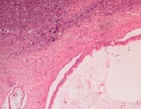
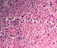
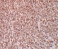
Mucinous tumors account approximately for 15% of all primary ovarian epithelial neoplasms. Ovarian mucinous tumors with mural nodules are rare surface epithelial-stromal tumorswhich can be of three types, "sarcoma-like"?, sarcoma or anaplastic carcinoma. Mural nodules of anaplastic carcinoma were first described in 1982 by Prat et al following which approximately fifty cases have been reported to date and in those cases, only one has been associated with benign mucinous tumors. The current case report is from a 50-year-oldpostmenopausal woman who presented to the gynecology outpatient department with complaints of abdominal pain and distension for the past five months. On histopathological examination, a diagnosis of Mucinous cystadenoma with focal atypia and mural nodule of anaplastic carcinoma was made and confirmed with immunohistochemistry.

Keywords: Anaplastic carcinoma, Ovarian mucinous neoplasms, Mural nodules

Corresponding Author How to Cite this Article To Browse
Lavanya M., Associate Professor, Department of Pathology, Sri Venkateshwaraa Medical College and Research Centre, Pondicherry, India.
Email:
Subashree K, Lavanya M, Jayanthi, Maria. Mural nodule of Anaplastic Carcinoma in an Ovarian Mucinous Cystadenoma- A rare case report. Trop J Pathol Microbiol. 2020;6(4):341-344.
Available From
https://pathology.medresearch.in/index.php/jopm/article/view/464

Introduction

Mucinous tumors account approximately for 15% of all primary ovarian epithelial neoplasmsand are only the second most common to serious tumors [1]. They can be classified into benign - mucinous cystadenomas, cystadenofibroma, or adenofibroma with focal low-grade atypia, borderline, and malignant- mucinous adenocarcinoma and cystadenocarcinoma.

Ovarian mucinous tumors may rarely harbor mural nodules which are also classified under surface epithelial-stromaltumors. The mural nodules can be of three types, benign "sarcoma-like"? and malignant sarcomatous nodules or anaplastic carcinomatous nodules [2]. They are known to be more associated with borderline tumors or carcinomas of intestinal-type [3]. Mural nodules of anaplastic carcinoma were first described in 1982 by Pratet al [4] following which only less than


Manuscript Received Review Round 1 Review Round 2 Review Round 3 Accepted
30-01-2020 07-02-2020 13-02-2020 19-02-2020
Conflict of Interest Funding Ethical Approval Plagiarism X-checker Note
No Nil Yes 17%

© 2020 by Subashree K., Lavanya M, Jayanthi, Maria and Published by Siddharth Health Research and Social Welfare Society. This is an Open Access article licensed under a Creative Commons Attribution 4.0 International License https://creativecommons.org/licenses/by/4.0/ unported [CC BY 4.0].

fifty cases have been reported to date. The occurrenceofa mural nodule of anaplastic carcinoma in benign mucinous cystadenoma makes it an even more rare diagnosis.

Case Report

A 50-year-oldpostmenopausal woman presented to the gynecology outpatient department with complaints of abdominal pain and distension for the past 5 months. Ultrasound abdomen showed loculatedascites with a left cystic ovarian neoplasm. Computed tomography abdomen showed a multiloculated, predominantly cystic lesion of the ovary with few solid components– possibly mucinous cystadenocarcinoma. The patient underwent staging laparotomy and the specimen was sent for histopathological examination. On gross examination, the left ovarian mass measured 25x20x14 cm. The external surface showed an intact capsule witha few congested blood vessels. Cut surface showed a multiloculated cyst filled with thick viscid mucinous secretions. Themulti location were measuringbetween 0.6 cm to 21 cm across. A careful examination led to the identification of multiple small areas of thickening (solid areas) in the cyst wall measuring between 0.2 to 0.5cm (<5% of the tumor). Tiny papillary projections were seen grossly in less than 5% of the areas. Uterus with the cervix, left and right fallopian tubes, right ovary, and omentum did not show any significant pathology. One right obturator, five right, andsix left external iliaclymph nodes identified were grossly free of tumor. Histopathology of the left ovarian mass showed cyst wall lined predominantly by endocervical type epithelium (columnar cells with a basally located nucleus and apical mucin vacuoles).The sections taken from the area of cyst wall with papillary projections showed nuclear stratification (2 to 3 layers) with micropapillary architecture and a moderate degree of pleomorphism in the lining epithelium. There was no evidence of invasion into the stroma. Sections studied from the solid areas showed denuded epithelium with underlying dispersed pleomorphic cells. Individual cells were large round to polygonal with abundant eosinophilic cytoplasm and peripherally placed vesicular nucleus with prominent nucleoli, resembling rhabdoid cells. Also seen were polygonal cells with scant cytoplasm and centrally placed irregular nucleus, few tumor giant cells with spindle (sarcomatoid) morphology, mitosis (6/10HPF) with few atypical forms, apoptotic bodies and focal areas of coagulative necrosis (Figure 1,2).

With these findings, we arrived at the diagnosis of Mucinous cystadenoma with focal atypia and a malignant mural nodule. Omentum, peritoneum, right and left external iliac, right obturator lymph nodes were free of tumor. Uterus with the cervix, left and right fallopian tubes and right ovary showed no significant pathology.

patho_464_01.jpg

Fig-1: Malignant stromal cells in a benign mucinous cyst. (H&E stain, 100X).

patho_464_02.jpg

Fig-2: Tumor cells with rhabdoid features (H&E stain, 400X).

Immunohistochemistry was performed in the solid areas of the left ovary to categorize the mural nodule. Vimentin showed cytoplasmic positivity and cytokeratin showed cytoplasmic positivity (Figure 3) in the lesional cells. CD 68 was negative.

patho_464_03.jpg

Fig-3: Cytokeratin Immunohistochemistry (100x). Tumor cells show positive staining.

Hence we arrived at a diagnosis of Mucinous cystadenoma with focal atypia and mural nodule of anaplastic carcinoma. There was no clarity if the current tumor had to be staged since the mucinous component was still benign. The postoperative period was uneventful, after which the patient was discharged and advised to be on regular follow up. The patient was not coming for a follow-up visit and


presented after three months to the oncology outpatient department with ascites. Imaging studies done showed peritoneal deposits and were advised chemotherapy. The patient again lost follow up and was found to have died after two months.

Discussion

Primary ovarian mucinous tumors include cystadenomas including cystadenomas with focallow-gradeatypia, mucinous borderline, or atypical proliferative mucinous tumors and carcinomas [5]. Mural nodules can occur in the entire spectrum butare seen to be more commonly associated withborderline tumors and carcinomas of intestinal-type. Mural nodules can be either reactive (sarcoma like a nodule) or malignant. Malignant nodules can show mesenchymal (sarcoma) or epithelial (anaplastic carcinoma) differentiation. The first case of the mural nodule was described in a mucinous tumor, in 1979, by Prat et al which was a sarcoma like a nodule and was described as a reactive lesion [6]. Following this, the same author also reported the mural nodule of sarcoma (1979) and anaplastic carcinoma (1982) [7]. The most commonly reported mural nodules are of carcinomatous types [8,9]. Sarcoma like mural nodulesis often multiple, circumscribed lesions, less than 5cm diameter [10]. On microscopy, they consist of osteoclast-like giant cells with pleomorphic spindle cells. Depending on the predominant cell type, three variants have been described- an epulis like nodule composed of multinucleated giant cells and mononuclear stromal cells; pleomorphic type composed of spindle cells and giant cells and the histiocytic type. The stroma appears edematous with hemorrhagic and necrotic foci [11]. Sarcoma like mural nodules is negative or weakly positive for Cytokeratin but positive for vimentin, CD68, and desmin [6]. Mural nodules of sarcoma are composed of malignant spindle cells that usually resemble fibrosarcoma or undifferentiated sarcoma [12,13]. The tumor cells are negative for cytokeratin and positive for vimentin. Intermediate filaments like act in or desmin are positive depending on the origin of sarcoma [6]. Mural nodules of anaplastic carcinomas usually occur in borderline tumorsor mucinous carcinomas with only one reported case associated with mucinous cystadenoma [14]. Mural nodule of anaplastic carcinoma may be confused on gross and microscopic examination with sarcoma and sarcoma like a nodule. It has been noted that most of the anaplastic carcinoma nodules are poorly

circumscribed with infiltrative borders, single and large (1-10cmin diameter) with stromal or vascular invasion and extensive areas of necrosis [11,15,16]. Some mural nodules are not grossly visible and are only identified microscopically. Mural nodules of anaplastic carcinoma are sub-classified microscopically into Rhabdoid type, composed of scattered polygonal cells with bright eosinophilic cytoplasm, eccentric nuclei, and prominent nucleoli; sarcomatoid type composed of sheets of spindle cells and the pleomorphic type which has both overlapping features.Pleomorphic type is the most commonly reportedmural nodule of anaplastic carcinoma. Mitotic figures are always present with few tumors showing less than 4 per 10 hpf. Immunohistochemistry for Cytokeratin and EMA is important for differentiating nodules of anaplastic carcinoma from other types. Both are positive in anaplastic carcinomatous nodules and are strong and diffuse in rhabdoid type. The tumor cells co-express vimentin in some cases. It is not clear if mural nodules are caused by cellular transformations within a single epithelial neoplasm or is a collision phenomenon of clonally unrelated tumors. But one case has shown KRAS expression in both the ovarian tumor and in the mural nodule component which represents a form of dedifferentiation. The current case is the second reported case of benign mucinous cystadenoma to be associated with a mural nodule of anaplastic carcinoma. This case grossly showed barely visible mural nodules of sizes less than 1cm with no obvious necrotic areas. Microscopically the tumor cells had overlapping features of rhabdoid and sarcomatoid cells. There were six mitotic figures per ten high power fields with focal areas of coagulative necrosis. The tumor cells were positive for vimentin and cytokeratin and negative for CD68. Stage IA tumors usually have a favorable prognosis with only occasional tumor-associated deaths. The prognosis of ruptured tumors or tumors which have spread beyond ovary usually has a bad prognosis [17]. Though this was a case of a mural nodule of anaplastic carcinomawhich occurred in a benign mucinous cyst the patient died in five months of diagnosis. Malignant mural nodules are fatal in 50% of cases and sarcoma like mural nodules though benign should be treated with caution [5].

Conclusion

This case has been chosen for discussion because of its extreme rarity and to emphasize the possibility of the malignant mural nodule in an otherwise


benign mucinous cystadenoma. Hence the need for careful gross and microscopic examination in all cases of ovarian mucinous cysts is mandatory.

Reference

  1. Provenza C, Young RH, Prat J. Anaplastic carcinoma in mucinous ovarian tumors- a clinicopathologic study of 34 cases emphasizing the crucial impact of stage on prognosis, their histologic spectrum, and overlap with sarcomalike mural nodules. Am J Surg Pathol. 2008;32(3)383-389.
  2. Mesbah Ardakani N, Giardina T, Amanuel B, Stewart CJ. Molecular profiling reveals a clonal relationship between ovarian mucinous tumors and corresponding mural carcinomatous nodules. Am J Surg Pathol. 2017;41(9)1261-1266.
  3. Allende DS, Drake RD, Chen L. Mural nodules of clear cell carcinoma in a mucinous borderline tumor of the ovary- a case report. Pathol Res Int. 2010.
  4. Prat J, Young RH, Scully RE. Ovarian mucinous tumors with foci of anaplastic carcinoma. Cancer. 1982;50(2)300-4.
  5. Bagué S, Rodríguez IM, Prat J. Sarcoma-like mural nodules in mucinous cystic tumors of the ovary revisited- a clinicopathologic analysis of 10 additional cases. Am J Surg Pathol. 2002;26(11)1467-1476.
  6. Bruijn JA, Smit VT, Que DG, Fleuren GJ. Immunohistology of a sarcomatous mural nodule in an ovarian mucinous cystadenocarcinoma. Int J Gynecol Pathol. 1987;6(3)287-293.
  1. Hillesheim PB, Farghaly H. Anaplastic spindle cell carcinoma, arising in a background of an ovarian mucinous cystic tumor- a case report with clinical follow up, review of the literature. Int J Clin Exp Pathol. 2010;3(8)808-811.
  2. Seidman JD,Cho KR,Ronnett BM,Kurman RJ. Blaustein's pathology of the female genital tract. 6th ed. New York- Springer, Chapter-14; Surface epithelial tumors of the ovary.
  3. Baergen RN, Rutgers JL. Mural nodules in common epithelial tumors of the ovary. Int J Gynecol Pathol. 1994;13(1)62-72.
  4. Chan YF, Ho HC, Yau SM, Ma L. Ovarian mucinous tumor with mural nodules of anaplastic carcinoma. Gynecol Oncol. 1989;35(1)112-119.
  5. Nichols GE, Mills SE, Ulbright TM, Czernobilsky B, Roth LM. Spindle cell mural nodules in cystic ovarian mucinous tumors- A clinicopathologic and immunohistochemical study of five cases. Am J Surg Pathol. 1991;15(11)1055-1062.
  6. Desouki MM, Khabele D, Crispens MA, Fadare O. Ovarian mucinous tumor with malignant mural nodules- dedifferentiation or collision?. Int J Gynecol Pathol. 2015;34(1)19-24.
  7. Kurman RJ, Carcangiu ML, Herrington CS, et al. WHO Classification of Tumours of Female Reproductive Organs. 4th ed, Lyon-IARC, Chapter 1-Mucinous tumors. 2014;p25-28.
  8. Hong SR, Chun YK, Kim YJ, Lim KT, Kim HS. Ovarian mucinous cystadenoma with mural nodule of anaplastic carcinoma. J Korean Med Sci. 1998;13(6)680-684.

  1. Baergen RN, Rutgers JL. Classification of mural nodules in common epithelial tumors of the ovary. Adv Anat Pathol. 1995;2;346-351.
  2. Zaloudek CJ, Garg K. Diagnostic Histopathology of tumors. 4th ed, Philadelphia- Saunders; Chapter 13- Tumors of the female genital tract. 2013; p669-675.
  3. Prat J, Scully RE. Sarcomas in ovarian mucinous tumors- A report of two cases. Cancer. 1979;44(4)1327-1231.