E-ISSN:2456-1487
P-ISSN:2456-9887
RNI:MPENG/2017/70771

Research Article

Swellings

Tropical Journal of Pathology and Microbiology

2020 Volume 6 Number 3 March
Publisherwww.medresearch.in

Observational study on FNAC evaluation of peripheral lymph node swellings

Raj P.1, Kumar P.2*
DOI: https://doi.org/10.17511/jopm.2020.i03.03

1 Pulak Raj, Consultant Pathologist, Nalanda Patho Centre, Biharsharif, Bihar, India.

2* Pawan Kumar, Professor and HOD, Department of Pathology, Vardhman Institute Of Medical Sciences, Pawapuri, Bihar, India.

Introduction: Peripheral lymphadenopathy is one of the common conditions encountered by clinicians. Knowledge of the pattern of diseases responsible for the same helps in better clinical decisions and management. The present study was conducted to assess the cytomorphological pattern of lymph node swellings by FNAC. Methods: The present study was cross-sectional in nature conducted upon 163 cases presenting with peripheral lymph node swellings for FNAC. Details of illness and findings of FNAC were noted. Results: Reactive hyperplasia was the most common finding in younger age groups (<20 years). Tuberculosis was more commonly seen in middle age groups (21-40 years). Malignant changes were more common in the elderly (>60 years). Conclusion: Reactive hyperplasia was the most common cause of lymphadenopathy in younger age groups was, tubercular lymphadenitis in the middle age group and metastatic carcinoma in patients above 60 years of age.

Keywords: Cytopathological changes, FNAC, Observational study, Peripheral lymphadenopathy

Corresponding Author How to Cite this Article To Browse
Pawan Kumar, Professor and HOD, Department of Pathology, Vardhman Institute Of Medical Sciences, Pawapuri, Bihar, India.
Email:
Raj P, Kumar P. Observational study on FNAC evaluation of peripheral lymph node swellings. Trop J Pathol Microbiol. 2020;6(3):223-229.
Available From
https://pathology.medresearch.in/index.php/jopm/article/view/443

Introduction

Lymph nodes are round, small nodules of lymphoid tissue that filter microorganisms and particulate matter from the lymph. It constitutes an important part of the defense system of the human body. Lymphadenopathy is the condition of enlargement of lymph nodes and is a common clinical presentation of patients consulting a clinician.

Lymph nodes are often involved in infectious diseases, many neoplastic conditions, lipid

storage diseases, endocrine disorders and various other conditions such as sarcoidosis and histiocytosis [1].

Surgical biopsy is considered to be the gold standard for diagnosis. However, it is costly, time-consuming and associated with more complications. Because, enlarged lymph nodes are easily accessible for aspiration, fine needle aspiration (FNAC) is advocated for the initial diagnosis and management of patients with lymphadenopathy [2].

The results can be available in a short time,


Manuscript Received Review Round 1 Review Round 2 Review Round 3 Accepted
13-02-2020 220-02-2020 27-02-2020 04-03-2020
Conflict of Interest Funding Ethical Approval Plagiarism X-checker Note
No Nil Yes 14%

© 2020 by Pulak Raj, Pawan Kumar and Published by Siddharth Health Research and Social Welfare Society. This is an Open Access article licensed under a Creative Commons Attribution 4.0 International License https://creativecommons.org/licenses/by/4.0/ unported [CC BY 4.0].

the technique being simple and leads to minimizing complications. It has been found that it offers a nearly accurate diagnosis of various pathologies including reactive lymphadenitis/inflammatory conditions, granulomatous disorders and neoplastic disorders. It identifies cases which need further investigations or follow-up. Hence, an excisional biopsy can be avoided in most of the cases [3]. De May has described the advantages of FNAC with the acronym SAFE. It means Simple, Accurate, Fast and Economical [4].

FNAC has a pivotal role in the evaluation of peripheral lymphadenopathy and can be used as an alternative to excision biopsy in developing countries with limited financial and health care resources [5]. The knowledge of the spectrum of lymphadenopathy in a particular area is important for making a definitive diagnosis [1].

Various researchers have described the pattern of diseases diagnosed on the basis of FNAC of peripheral lymph nodes in different parts of the country [6-13]. Very few studies regarding this have been done in this area. Hence, this study was conducted.

Aims and objectives- The present study was conducted to assess the cytomorphological pattern of lymph node swellings by FNAC in patients presenting with peripheral lymph node swellings.

Material and Methods

Study setting: The present study was conducted at Nalanda Patho Centre, Biharsharif, Bihar. It is a specialized pathological laboratory providing services to patients of Biharsharif and neighbouring districts of Bihar for the past 30 years and has all the modern diagnostic equipment.

Duration and type of study: The present study was cross-sectional in nature conducted between November 2019 to January 2020.

Study subjects: Study subjects included patients reporting to the laboratory for FNAC who was suffering from peripheral lymph node swellings. A total of 163 cases were studied.

Inclusion criteria: Patients above 18 years of age suffering from peripheral lymph node swellings who underwent FNAC were included in the present study.

Exclusion criteria: Seriously ill cases, those who did not give consent or in which FNAC was contraindicated were excluded.

Sampling: All the patients with peripheral lymph node swellings reporting during the study period and fulfilling the selection criteria were included in the present study. A total of 163cases were included.

Data collection procedure: The patients were cases with peripheral lymph node swellings referred to the laboratory by the clinicians for FNAC. History was taken from the patients and findings were noted. The patients were explained about the procedure and informed consent was taken from them.

FNAC was done with aseptic precautions using 10 cc disposable syringe and 22/23-gauge needle by both aspiration and non-aspiration technique as required. Smears were prepared on two slides. One of the slides was air-dried and stained with May-Grunwald Giemsa stain.

Another slide was wet fixed with alcohol and was stained with Papanicolaou stain. This was followed by careful microscopic examination and the cytological diagnosis was done. A pre-tested semi-structured proforma was used for noting the findings.

Data analysis: Data was entered in Microsoft Excel 2013 and data analysis was done using Statistical Package for Social Sciences (SPSS) v 16.0. Data were summarized as frequency and percentage for categorical variables and mean and SD for numerical variables. Statistical tests were done as needed. the p-value of <0.05 was considered to be statistically significant.

Ethical consideration and permission: Approval from the Institutional Ethics Committee was obtained. The participants were informed about the study and informed consent was taken. The confidentiality of records was maintained.

Results

The present study included 163cases coming for FNAC of peripheral lymph node swellings. The age of the patients ranged from 2 to 79 years. Most of the cases belonged to the age group of 21-30 years. There were 63.2% males and 36.8% females resulting in male: female ratio of 1.72:1.

Table-1 shows the common sites of lymphadenopathy. Most of the cases presented with cervical lymphadenopathy. Generalized lymphadenopathy was seen in 1.2% of cases.


Table-1: Site of peripheral lymphadenopathy.1

Site Frequency %
Cervical 133 81.6
Axillary 19 11.7
Inguinal 9 5.5
Generalized 2 1.2
Total 163 100

Table-2 shows the cytological diagnosis of the cases. Tubercular lymphadenitis was the most common finding in 41.1% of cases followed by reactive hyperplasia (27.6%). Metastatic carcinoma was seen in 9.8% of cases.

Table-2: Cytological diagnosis.

Cytological diagnosis Frequency %
Acute lymphadenitis 27 16.6
Chronic reactive hyperplasia 45 27.6
Tuberculous lymphadenitis 67 41.1
Metastatic carcinoma 16 9.8
Lymphoma 8 4.9
Total 163 100

Table-3 shows the cytomorphological findings in different age groups. Reactive hyperplasia was the most common finding in younger age groups (<20 years). Tuberculosis was more commonly seen in middle age groups (21-40 years). Malignant changes were more common in the elderly (>60 years).

Table-3: Cytomorphological findings in different age groups.

Age group Acute lymphadenitis Reactive hyperplasia tuberculosis Hodgkin’s Lymphoma NHL Metastasis Total
<10 8 8 4 0 0 0 20
11-20 7 16 8 1 0 0 32
21-30 6 7 25 0 0 0 38
31-40 4 6 12 0 0 1 23
41-50 2 3 11 2 2 4 24
51-60 0 4 5 0 2 8 19
>60 0 1 2 0 1 3 7
Total 27 45 67 3 5 16 163

Discussion

Lymphadenopathy is one of the common conditions encountered in outpatients. It is a clinical manifestation of regional or systemic diseases and gives a clue to the underlying disease. It can be due to benign or malignant causes. The etiology varies according to the geographical condition. It becomes important to identify the underlying cause for proper diagnosis and adequate management. Excision biopsy is a gold standard diagnostic test but is associated with complications.

FNAC has been established as a safe, cheap and reliable test for diagnosis of superficial masses. It is used as the first-line investigation in the initial management of lymphadenopathy cases and has been advocated as a useful method in comparison with more expensive surgical excision biopsies, especially in developing countries with limited resources.

Awareness about the prevailing pattern of causes of lymphadenopathy in a particular area makes the task of clinician easier. Hence, the present study was conducted to assess the cytomorphological

pattern of lymph node swellings by FNAC in patients presenting with peripheral lymph node swellings.

A total of 163 cases with peripheral lymphadenopathy reporting for FNAC were included in the present study. History was taken from the patients and findings were noted. The patients were explained about the procedure and informed consent was taken from them. FNAC was done as per guidelines under aseptic precautions and slides were prepared. Microscopic examination was done and findings were noted.

In two cases, adequate material could not be obtained and 161 slides (98.8%) were examined. Malhotra et al also reported that 1.7% of cases yielded inadequate material.

Cytomorphological features were used for diagnosis of pathology as discussed by Shah et al [14]. Granulomatous lymphadenitis was diagnosed by the presence of epithelioid cell granulomas (Figure-1) in Giemsa stained smear at 400X magnification.

Reactive lymph node hyperplasia was diagnosed by observing the polymorphous population of lymphoid cells composed of centrocytes, centroblasts, small


lymphocytes, and immunoblasts. Suppurative lymphadenitis was diagnosed by the presence of sheets of neutrophils. Necrotic lymphadenitis was marked by extensive areas of necrosis. Ziehl-Nelson stain was used for finding AFB.

Tuberculosis was diagnosed when AFB was positive. Microfilaria was also searched for in lymph node aspirate. Non-Hodgkin's lymphoma was diagnosed by the presence of a monomorphic population of lymphocytes scattered singly in a highly cellular smear and the absence of RS cell.

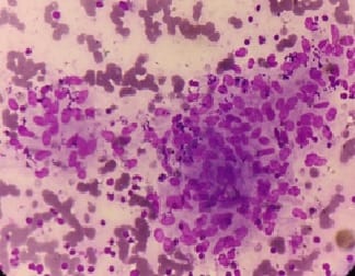
Diagnosis of Hodgkin’s lymphoma (Figure-2) was made on the basis of the presence of either mononuclear Hodgkin’s cells or Classical RS cells in the background of lymphocytes, eosinophils, plasma cells, and histiocytes. Metastatic deposits were diagnosed by the presence of cytological patterns and cellular details.

Age and sex- In the present study, the age of the patients ranged from 2 to 79 years. Most of the cases belonged to the age group of 21-30 years. There were 63.2% males and 36.8% females resulting in male: female ratio of 1.72:1.

In the study done by Badge et al, it was seen that age of the patients ranged from 2.5 to 87 years. The youngest patient was diagnosed as tubercular lymphadenitis, and the oldest patient was diagnosed as having metastatic deposits in the lymph node from a squamous cell carcinoma. The majority (74.3%) of the patients were in the age group of 11–50 years, with a peak in the age group 21-30 years. Only 15 patients were aged <10 years and one aged above 80 years. The male to female ratio was 1:1.64 [5].

Sharma et al also found that the ages of patients were from 6 months to 65 years in which 54% were males and 46% were females. The youngest patient with lymphadenopathy was diagnosed as Suppurative lymphadenitis and the oldest patient was diagnosed with metastatic adenocarcinoma [15]. Farooq et al reported that the mean age of the patients was found to be 32.55 years.

Most of the patients (41%) belonged to the age group 15-45 years [3]. Malhotra et al found similar results. The mean age of all the patients presenting with lymphadenopathy was 21.67 years. The male-to-female ratio of 1.2:1 was observed, with males contributing 54.7% cases and females 45.29% cases. Shrivastav et al divided cases into three groups depending on their age: Group I (0-20

years), Group II including adolescent and middle- aged patients (21-50 years) and Group III with elderly patients (>51 years).

In Group I, there was 36.98% patients with a male: female ratio of 1.22:1; in Group II total 49.11% cases and male: female ratio was 0.92:1; in Group III there were 13.90% cases with a male: female ratio of 2.55:1. Shah et al found that the range of age of patients was from 5 months to 90 years in which 60.2% were males and 39.8% were females. Farooq et al reported that 58% were males and 42% were females [4].

Site and Size- Most of the cases presented with cervical lymphadenopathy (81.6%). Generalized lymphadenopathy was seen in 1.2% of cases. Lymph nodes of > 3cm size were seen in 19.1% cases.

Shrivastav et al found that among the various sites of lymph node involvements, cervical lymph nodes were the most common (85.82%) cases followed by, supraclavicular (4.22%), inguinal nodes (3.48%), axillary (2.73%) and in (3.73%) of cases more than 1 site was involved [4].

Shah et al observed that among the various sites of lymph node involvement, maximum numbers were of the cervical region (88.5%), followed by axillary region (6.5%) and inguinal region (4.1%). 0.9% of cases showed generalized lymphadenopathy [14].

The most common site of involvement was cervical lymph nodes comprising the majority (32%) of the cases in the study conducted by Farooq et al [3]. Sharma et al noted that cervical lymph nodes were involved most commonly (84%) followed by axillary (3%), inguinal (2%) and generalized (1%) [15].

Malhotra et al observed that 39.73% of patients had solitary lymphadenopathy and 135 62.26% of patients had multiple enlarged lymph nodes. The cervical region was found to be the most common site of lymphadenopathy (71.79%) followed by axillary region (11.11%), inguinal region (6.41%), abdominal region (4.70%) and mediastinal region (1.79%).

Deep‑seated lesions were aspirated under image guidance (both computed tomography and ultrasonography). Of the 168 patients with cervical lymphadenopathy, 26.19% had sub‑mandibular lymph nodes, 9.52% had supraclavicular, and 3.57% had submental lymph nodes. The size of the lymph nodes was <1 cm in 6.41% cases, ranging from 1 to 3 cm in 78.20% and >3 cm in the


remaining 14.95% [1].

Diagnosis- In the present study, tubercular lymphadenitis was the most common finding in 41.1% of cases followed by reactive hyperplasia (27.6%). Metastatic carcinoma was seen in 9.8% of cases. Reactive hyperplasia was the most common finding in younger age groups (<20 years). Tuberculosis was more commonly seen in middle age groups (21-40 years). Malignant changes were more common in the elderly (>60 years).

Badge et al also found tubercular lymphadenitis in 48.70% cases followed by granulomatous lymphadenitis in 32.90% cases, reactive hyperplasia in 8.06% cases, metastatic carcinoma in 3.87% cases, suppurative granulomatous in 2.25% cases, acute suppurative lymphadenitis in 1.61% cases, non‑Hodgkin’s lymphoma in 0.64% cases and Hodgkin’s lymphoma in 0.32% case. Metastatic deposits were more common in the elderly age group (greater than the sixth decade). Tuberculous and granulomatous lymphadenopathies were more common in the third to a fourth decade [5].

Sharma et al observed that granulomatous lymphadenitis was the most common followed by reactive lymphadenopathy, Suppurative –necrotic lesions, micro filarial lymphadenitis, tuberculosis. Among the neoplastic lymph node lesions, the commonest was metastasis followed by lymphoma. Metastatic deposits were more common in the extremes of ages. Tuberculosis and granulomatous pathology were more common in the 10-40years [15].

In the study done by Farooq et al, 37% of cases were diagnosed as reactive lymphadenitis, 32% as tubercular lymphadenitis, 4% as suppurative lymphadenitis, and 17% as metastatic while 5% cases as Hodgkin’s lymphoma and 5% cases as Non-Hodgkin’s lymphoma. Tuberculous lymphadenitis was found to be the most common cause of lymphadenopathy in this age group (46.34%) followed by reactive lymphadenitis (36.58%). 3 out of 5 cases (60%) of Hodgkin’s lymphoma belonged to this age group.

In the pediatric age group (0-14 years), the most common cause of lymphadenopathy was found to be reactive lymphadenitis (51.72%) followed by tuberculous lymphadenitis (27.59%). In the elderly age group (> 45 years), metastatic carcinoma was found to be the most common cause of lymphadenopathy (43.33%). Out of 5 cases of Non-Hodgkin’s lymphoma 4 i.e. 80% belonged to this

age group.

The most common cause of lymphadenopathy in males was found to be reactive hyperplasia (37.93%) followed by tuberculous lymphadenitis (27.59%). Similarly, in females most common cause of lymphadenopathy was found to be tuberculous lymphadenitis (38.09%) followed by reactive hyperplasia (35.71%). Out of 17 secondary metastatic cases, 11 were males and 6 were females [3].

Shah et al found that 66.9% cases were of infective etiology, 31.3% were of metastatic carcinoma and 1.8% cases were of lymphoma. In infective etiology, the maximum number of cases were of tuberculous lymphadenitis followed by chronic reactive hyperplasia. Tuberculous lymphadenitis was found to be the most common pathologic lesion accounting for 44.8% of cases followed by metastatic malignancy constituting 31.3% of cases.

Chronic reactive hyperplasia was seen most often (69.9%) in the first three decades, tuberculous lymphadenitis (73.6%) in the second and third decades. Metastatic carcinoma was seen 81.3% after the age of 40 years.

Cases of lymphoma were distributed in all age groups. Male predominance was noted in acute lymphadenitis, metastatic carcinoma and malignant lymphoma with a male: female ratio of 1.1:1, 4:1, 4:1 respectively while in reactive lymphadenitis and tuberculous lymphadenitis there was equal distribution with a male: female ratio is 1:1 [14].

Shrivastav et al observed that in each of the three groups, the most common cause of lymphadenopathy was different. In Group I, the most frequent cause of lymphadenopathy was found to be reactive lymphoid hyperplasia with 52.38% cases followed by tubercular lymphadenitis in 39.41% cases.

In Group II, the most common etiology of lymphadenopathy was found to be tubercular (46.21%) followed by reactive lymphoid hyperplasia with 28.08% cases.

Metastasis was observed as the most frequent causative agent of lymphadenopathy with 56.33% cases in Group III, followed by tubercular lymphadenitis in 16.30% cases. Out of total cases of metastatic carcinoma, squamous cell variety was predominant (65%) followed by adenocarcinoma (23%), 2% cases having small cell carcinoma and 10% cases having undifferentiated carcinoma [4].


patho_443_01.jpg

Fig-1: Granulomatous lymphadenitis using MGG stain (400X).

patho_443_02.jpg

Fig-2: Hodgkin’s lymphoma using MGG stain (400X).

Conclusion

It is concluded from the present study that the etiology of lymphadenopathy was different in different age groups. Reactive hyperplasia was the most common cause of lymphadenopathy in younger age groups, tubercular lymphadenitis in the middle age group and metastatic carcinoma in patients above 60 years of age. As there is the high rate of malignancy in the elderly, special precaution should be taken in doing FNAC in these patients in terms of preparation of more of wet fixed smears and keeping unstained smears which might be needed for further staining and studies. FNAC can be a reliable and convenient screening tool for the first-line diagnosis. For the cases in which diagnosis

is not confirmed, the further histological examination can be done for definitive treatment of the cases.

Limitations of the study- The present study is observational in nature and only describes the clinical and cytological profile of peripheral lymph node swellings. Further study is needed to compare these findings with biopsy to assess the diagnostic value of FNAC.

What does the study add to the existing knowledge?

The findings of FNAC of peripheral lymph node swellings in this area have been described. The patient profile and pathological changes have been elaborated.

Author’s contribution

Dr. Pulak Raj: Principal author collected data and has written the report.

Dr. Pawan Kumar Chaudhary: Edited the article, did a review of the literature and has written the discussion.

Reference

  1. Shrivastav A, Shah H, Agarwal N, Santwani P, Srivastava G. Evaluation of peripheral lymphadenopathy by fine needle aspiration cytology- A three-year study at tertiary center. J NTR Univ Health Sci. 2014;3(2)86-91.
  2. Parthy P, Hota S, Dash S, Samartaray S, Panda S, Rout N. Analysis of FNAC in diagnosis of lymphadenopathy –a retrospective study from a regional cancer centre, Cuttuck, Odisha. Int J Res Med sci. 2017;5(12)5287-5292.
  3. K P SK, G S S, Ahmed SM. A prospective study of cervical lymphade nopathy in 78 children in tertiary hospital- in Kerala. JEMDS. 2015;4(33)5617-5629.

  1. Malhotra A, Lahori M, Nigam A, Khajuria A. Profile of lymphadenopathy- An institutional based cytomorphological study. Int J App Basic Med Res. 2017;7(2)100-103.
  2. Farooq S, Amain J, Singh K, Jahan S. Evaluation of Lymphadenopathy using FNAC as a Diagnostic Tool. Int J Contemp Med Res. 2018;5(1)24-27.
  3. Badge S, Ovhal A, Azad K, Meshram A. Study of fine-needle aspiration cytology of lymph node in rural area of Bastar District, Chhattisgarh. Med J DY Patil Univ. 2017;10(2)143-148.
  4. Duraiswami R, Sujatha M, Chandran P, Prakash A. Spectrum of pathologies on FNAC evaluation of peripheral lymph nodes at a tertiary care center in Hyderabad- a retrospective study. Int J Adv Med. 2017;4(1)27-33.
  5. Pavithra P, Geetha J. Role of fine needle aspiration cytology in the evaluation of the spectrum of lymph node lesions. Int J Pharm Bio Sci. 2014;5(4)377-384.
  6. Shah PC, Patel CB, Bhagat V, Modi H. Evaluation of peripheral lymphadenopathy by fine needle aspiration cytology- a one-year study at tertiary centre. Int J Res Med Sci. 2016;4(1)120-125.
  1. Janagam C, Atla B. Role of FNAC in the diagnosis of cervical lymphadenopathy. Int j res med sci. 2017;5(12)5237-5241.
  2. Khajuria R, Goswami K, Singh K, DUbey V. Pattern of Lymphadenopathy on Fine needle aspiration cytology in Jammu. JK Sci. 2006;8(3)157-159.
  3. Mishra B, Sinha A, Singh S. A study of Patterns and etiologies of peripheral lymphadenopathy in a tertiary care hospital in Ranchi, India. IOSR Journal of Dental and Medical Sciences. 2017;16(5)46-50.
  4. Paikrao Y, Chintale K. Study of diagnosis of tuberculosis cervical lymphadenopathy by FNAC in rural tertiary health care centre. Int J App Res. 2017;3(12)96-101.
  5. Sharma BH, Bansal M, Kumar N, Gupta M. Spectrum of lymphadenopathy on fine needle aspiration cytology-A 3 year retrospective study in a tertiary care centre. ACHR. 2019;4(1)82-86.
  6. Sheela K, Priya M. Reliability of FNAC as a diagnostic tool in lymphadenopathy. Int J Adv in Med. 2017;4(4)1073-1077.