E-ISSN:2456-1487
P-ISSN:2456-9887
RNI:MPENG/2017/70771

Research Article

Microbiological

Tropical Journal of Pathology and Microbiology

2020 Volume 6 Number 2 February
Publisherwww.medresearch.in

Incidence, microbiological profile and outcome of ventilator-associated events in a tertiary care hospital in Bangalore, Karnataka, India

Shashikala N.1, Manasa S.2*, Hedge D.3, Vikram V.4, Kumar P M.5
DOI: https://doi.org/10.17511/jopm.2020.i02.04

1 Shashikala N, Infection Control Officer, Department of Pathology, Sagar Hospital, Bangalore, Karnataka, India.

2* Manasa S, Junior Infection Control Officer, Department of Pathology, Sagar Hospital, Bangalore, Karnataka, India.

3 Deviprasad Hedge, Head of the Department (ICU), Department of Pathology, Sagar Hospital, Bangalore, Karnataka, India.

4 Venkatesh Vikram, Medical Director, Department of Pathology, Sagar Hospital, DSI, Bangalore, Karnataka, India.

5 Mahendra Kumar P, Medical Director, Department of Pathology, Sagar Hospital, Bangalore, Karnataka, India.

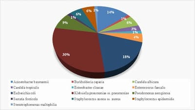
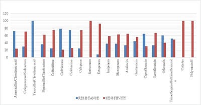
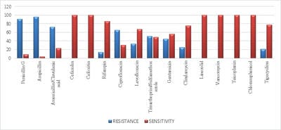
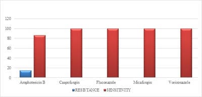
Introduction: Despite different ICU admission causes, ventilator-associated event (VAE) is still a common cause of mortality and morbidity in intubated patients and impedes obvious progression in diagnostic modalities and management of these infections. Objectives: The aim of this study was to estimate VAE incidence, detection of antimicrobial resistance pattern’s which can help in the management and to follow the outcome of the patients having VAE. Materials and methods: This was a retrospective study done over a period of 24months from January 2018 to December 2019 in a tertiary care hospital in Bangalore. The study population included all the confirmed VAE patients. Their incidence of microbiological profile and the outcome was noted. Results: Out of 422 mechanical ventilated (MV) patients 71 patients developed VAE. In the 71 proved VAE cases 48 (67.6%) were male and23 (32.4%) were females. Out of 71 cases, the mean age who were having VAE falls in the range of 65- 75 yrs. The incidence of VAE is 16.82 and the incidence density:9.78 /1000 ventilator days. Klebsiella pneumonia (29.5%) was the common organism isolated among 71 cases. Conclusion: Utilizing the preventive, diagnostic, and treatment recommendations outlined should allow for improved outcomes for a common and serious medical complication seen in ICU mechanically ventilated patients.

Keywords: Ventilator-associated event, Ventilator-associated event pneumonia, Mechanical ventilation

Corresponding Author How to Cite this Article To Browse
Manasa S, Junior Infection Control Officer, Department of Pathology, Sagar Hospital, Bangalore, Karnataka, India.
Email:
Shashikala N, Manasa S, Hedge D, Vikram V, Mahendra KP. Incidence, microbiological profile and outcome of ventilator-associated events in a tertiary care hospital in Bangalore, Karnataka, India. Trop J Pathol Microbiol. 2020;6(2):130-138.
Available From
https://pathology.medresearch.in/index.php/jopm/article/view/420

Manuscript Received Review Round 1 Review Round 2 Review Round 3 Accepted
28-01-2020 07-02-2020 14-02-2020 19-02-2020
Conflict of Interest Funding Ethical Approval Plagiarism X-checker Note
No Nil Yes 18%

© 2020 by Shashikala N, Manasa S, Deviprasad Hedge, Venkatesh Vikram, Mahendra Kumar. P and Published by Siddharth Health Research and Social Welfare Society. This is an Open Access article licensed under a Creative Commons Attribution 4.0 International License https://creativecommons.org/licenses/by/4.0/ unported [CC BY 4.0].

Introduction

Although invasive mechanical ventilation is not easy to take a decision for some patients, it is a method of keeping a patient alive by adequate tissue oxygenation to support the body during the treatment course in the ICU [1].

Patients are admitted to the ICU for many reasons either related to pulmonary diseases such as acute respiratory failure and massive pneumonia or other causes such as neuromuscular diseases, after major surgeries, shock, or post-arrest [2].

Invasive mechanical ventilation (MV) is used as a cornerstone in the treatment plan of these patients [3]. Despite the different ICU admission causes, the mortality rates in intubated patients are still higher than those who do not need ventilator support [4].

Ventilator-associated event pneumonia is pneumonia that develops 2–3 days after endotracheal intubation; the patient must have new or progressive radiological infiltrate, infection alerts (e.g. fever, white blood cell count change), altered sputum characters, and isolation of a causative organism, all together to diagnose VAE [5].

VAE is still one of the most common hospital-acquired infections that are encountered in ICU patients despite the recent progress in diagnostic modalities and management advances of these infections [6].

Pneumonia is usually mild or low in severity if it occurs in the early period of invasive ventilation and the organisms are most responsive to the antibiotics administered Whereas after a few days (late-onset), pneumonia is more severe in its course, with fewer organisms responding to antibiotics and increased rate of morbidity and mortality among those with late-onset infection [7].

Materials and Methods

Source of data: This is a retrospective study done over a period of 24months from January 2018 to December 2019. The study will be conducted in the Department of Microbiology, in a tertiary care hospital.

Inclusion criteria

All the adult patients who were on mechanical ventilation in ICU were included in the study. In those patients who had VAE were identified.

Exclusion criteria

  1. ICU patients whose hospital stay was shorter than 2 days
  2. patients who showed symptoms of infection within 2 days of MV
  3. Patients who died or developed pneumonia within 48 h,
  4. Patients on high-frequency ventilation,
  5. Patients on extracorporeal life support or Para corporeal membrane oxygenation
  6. Neonates

were excluded from the study.

Methodology: The present study retrospectively reviewed all the adult cases who were intubated and under mechanical ventilation in ICU and diagnosed the VAE in them by standard criteria according to the recent guidelines by CDC. Different specimens were collected from all the mechanically ventilated patients. Specimens were cultured according to the standard microbiological standards and their sensitivity to drugs and also their outcome was studied in detail.

Table-1: Threshold value for culture specimens

Specimen collection /technique Values
Lung tissue ≥ 104 cfu/g tissue*
Bronchoscopically (B) obtained specimens
Bronchoalveolar lavage (B-BAL) ≥ 104 cfu/ml*
Protected BAL (B-PBAL) ≥ 104 cfu/ml*
Protected specimen brushing (B-PSB) ≥ 103 cfu/ml*
Nonbronchoscopically (NB) obtained (blind) specimens
NB-BAL ≥104 cfu/ml*
NB-PSB ≥ 103 cfu/ml*
Endotracheal aspirate (ETA) ≥ 105 cfu/ml*

Results

The medical charts were reviewed for demographic (age, sex) data, clinical and laboratory data of VAE patients were also collected. Our records search showed around 422 intubated cases in ICU in those patients 71 patients had developed VAE. All the71 cases fulfilled the diagnostic criteria for VAE. In the 71 proved VAE cases 48(67.6%) were male and23 (32.4%) were females. Out of 71 cases, the mean age who were having VAE falls in the range of 65- 75 yrs. In terms of the development of VAE in relation to the duration of ventilation, 42 (59.2%) patients developed VAE during 15 days or more of MV, whereas 29(40.8%) patients developed VAE


during less than 15days of MV.

The different specimen was collected from the suspected patients like BAL fluid, endotracheal aspirate, etc. Those specimens were cultured and their sensitivity was also tested using standard microbiological procedures. The most common organisms isolated were Klebsiella pneumoniae ss. Pneumoniae 21(29.5%) followed by Escherichia coli 13 (18.3%), Acinetobacter baumanni 10 (12.6%), Pseudomonas aeruginosa 6 (8.4%). The other organisms isolated were Staphylococcus aureus, Staphylococcus epidermidis and Candida albicans 4 (5.6%) each. A single 1(1.4%) isolates of Burkholderia cepacia, Stenotrophomonas maltophilia, Serratia fonticola were also seen. The isolates have been depicted in Figure 1.

patho_420_01.jpg

Fig-1: Microbiological profile of VAP cases.

The sensitivity of the organisms isolated was done by standard microbiological procedures. In gram-negative microorganisms isolated, any resistance to Colistin and polymyxin B was not observed but carbapenem resistance was noted. In gram-positive organisms, no vancomycin-resistant species were seen and in fungal isolates, all were sensitive to caspofungin, fluconazole, variconazole, and micafungin. This is depicted in Figure 2, Figure 3 and Figure 4 as follows.

patho_420_02.jpg

Fig-2: Antibiogram of gram-negative organisms

patho_420_03.jpg

Fig-3: Antibiogram of gram-positive organisms.

patho_420_04.jpg

Fig-4: Sensitivity pattern of antifungals agents

Discussion

Previously, the diagnosis of VAE is based on a combination of clinical, radiological, and microbiological criteria. There was a wide range of clinical conditions that mimic VAE in ventilated patients, including acute respiratory distress syndrome (ARDS), pulmonary edema, pulmonary contusion, tracheobronchitis, and thromboembolic disease. Some of the clinical features used to define a VAE (e.g. change in tracheal secretions) are subjective and are subject to inter- and intra-observer variation. The diagnostic value of these clinical criteria in isolation, and in combination, was reviewed recently by Klompas [8]. While individual clinical criteria appear to lack clinical sensitivity, a combination of clinical criteria with laboratory criteria and radiological features improves the accuracy of clinical diagnosis [8]. Fabregas and colleagues found radiological infiltrate plus two from three of fever, leucocytosis, and purulent secretions, to have a sensitivity of 69% and specificity of 75% for diagnosing VAE. [9].

There are no radiological criteria pathognomonic of VAE and the interpretation of chest radiographs in ventilated patients is very difficult. Single air bronchograms and fissure abutment are highly specific, but they lack sensitivity [8]. Invasive and non-invasive sampling techniques are used to obtain microbiological specimens to diagnose VAE.


Invasive techniques include bronchoscopic alveolar lavage (BAL) and protected specimen brushings (PSB), while less invasive techniques include mini BALs. Tracheal aspirates are the least invasive to obtain but the most likely to be contaminated with oro-pharyngeal colonizing bacteria. Quantitative cultures are often used to differentiate between colonization and infection. The diagnostic threshold for BALs is 104 colony-forming units per milliliter (CFU ml−1) and this is often the gold standard against which other diagnostic criteria are compared. However, as bronchoscopic sampling cannot guarantee sampling from the area of the lung most affected, the sensitivity of this test is low, although the specificity is quite high (significant false-negative rate). Several studies have compared the value of quantitative invasivevsnon-quantitative, non-invasive cultures. Meta-analyses comparing these have come to the conclusion that neither method confers any advantage on survival, length of ICU stay, or duration of mechanical ventilation [10].

Definitions: Previously The Clinical Pulmonary Infection Score (CPIS) was developed by Pugin and colleagues to facilitate the diagnosis of VAE using clinical variables [11]. It gives a score of 0–3 for temperature, leucocytosis, ratio, chest radiography, tracheal secretions, and culture of tracheal aspirate. The maximum score that can be obtained is 12 and a score >6 is diagnostic of VAP. The assessment of the CPIS score is prone to considerable inter-

observer variability, particularly with regard to the interpretation of the tracheal secretions and the chest X-ray (CXR).

The Centre for Disease Control (CDC) definition was designed as a surveillance tool for HAI but has been used in the diagnosis of VAE [12]. However, it has been shown to have good sensitivity and positive predictive value, but its low specificity limits its value when compared with bronchoscopic cultures.

The Johannson criteria diagnosed a VAE based on the presence of new or progressive infiltrates on the CXR associated with at least two of three clinical features—leucocytosis, purulent secretions, temperature >38°C. Diagnosis by these criteria was compared with immediate post-mortem lung biopsies; the sensitivity was only 69%, while the maximum specificity was 75% [9].

The HELICS criteria were used for VAE surveillance in Europe [10]. These again rely on a combination of clinical, radiological, and microbiological criteria and classify pneumonia from PN1 to PN5 based on the microbiological method used. PN1 refers to diagnosis by minimally contaminated lower respiratory tract (LRT) specimens (BAL, PBS, distal protected aspirates), while PN4 refers to positive sputum culture or to non-quantitative LRT aspirates such as tracheal aspirates. Therefore, a unit's VAE rate can vary significantly depending on the microbiological method used.


Table-1: Current Definition of VAE according to CDC 2020 guidelines

Ventilator-associated condition (VAC) The patient has a baseline period of stability or improvement on the ventilator, defined by ≥ 2 calendar days of stable or decreasing daily minimum* FiO2 or PEEP values. The baseline period is defined as the 2 calendar days immediately preceding the first day of increased daily minimum PEEP or FiO2. *Daily minimum defined by the lowest value of FiO2 or PEEP during a calendar day that is maintained for > 1 hour. After a period of stability or improvement on the ventilator, the patient has at least one of the following indicators of worsening oxygenation: 1) Increase in daily minimum* FiO2 of ≥ 0.20 (20 points) over the daily minimum FiO2 of the first day in the baseline period, sustained for ≥ 2 calendar days.2) Increase in daily minimum* PEEP values of ≥ 3 cmH2O over the daily minimum PEEP of the first day in the baseline period†, sustained for ≥ 2calendar days.*Daily the minimum defined by the lowest value of FiO2 or PEEP during a calendar day that is maintained for > 1 hour. Daily minimum PEEP values of 0-5 cmH2O are considered equivalent for the purposes of VAE surveillance.
Infection-related Ventilator-Associated Complication (IVAC) On or after calendar day 3 of mechanical ventilation and within 2 calendar days before or after the onset of worsening oxygenation, the patient meets both of the following criteria:1)Temperature > 38 °C or < 36°C, OR white blood cell count ≥ 12,000 cells/mm3 or ≤ 4,000 cells/mm3.AND2)A new antimicrobial agent(s) (see Appendix for eligible antimicrobial agents) is started, and is continued for ≥ 4 qualifying antimicrobial days(QAD).

Possible Ventilator-Associated Pneumonia (PVAP) On or after calendar day 3 of mechanical ventilation and within 2 calendar days before or after the onset of worsening oxygenation, ONE of the following criteria is met (taking into account organism exclusions specified in the protocol): Criterion 1: Positive culture of one of the following specimens, meeting quantitative or semi-quantitative thresholds† as outlined in protocol, without requirement for purulent respiratory secretions: Endotracheal aspirate, ≥ 105 CFU/ml or corresponding semi-quantitative result, Bronchoalveolar lavage, ≥ 104 CFU/ml or corresponding semi-quantitative result, Lung tissue, ≥ 104 CFU/g or corresponding semi-quantitative result, Protected specimen brush, ≥ 103 CFU/ml or corresponding semi-quantitative result. Criterion 2: Purulent respiratory secretions (defined as secretions from the lungs, bronchi, or trachea that contain >25 neutrophils and <10 squamous epithelial cells per low power field [lpf, x100])† PLUS organism identified from one of the following specimens (to include qualitative culture or quantitative/semi-quantitative culture without sufficient growth to meet criterion #1): Sputum, Endotracheal aspirate, Bronchoalveolar lavage, Lung tissue, Protected specimen brush. Criterion 3: One of the following positive tests: Organism identified from pleural fluid (where specimen was obtained during thoracentesis or initial placement of chest tube and NOT from an indwelling chest tube)Lung histopathology, defined as 1) abscess formation
  or foci of consolidation with intense neutrophil accumulation in bronchioles and alveoli; 2) evidence of lung parenchyma invasion by fungi (hyphae, pseudohyphae or yeast forms); 3) evidence of infection with the viral pathogens listed below based on results of immunohistochemical assays, cytology, or microscopy performed on lung tissue. Diagnostic test for Legionella species, Diagnostic test on respiratory secretions for influenza virus, respiratory syncytial virus, adenovirus, parainfluenza virus, rhinovirus, human metapneumovirus, coronavirus.

VAE Pathogenesis

The main pathogenic factor in the development of VAE is biofilm formation within the tracheal tube (TT) and microaspiration of secretions. The presence of a TT interferes with the normal protective upper airway reflexes and prevents effective coughing. The oropharynx becomes rapidly colonized by aerobic gram-negative bacteria after illness, antibiotic administration, and hospital admission. These contaminated secretions pool above the TT cuff and slowly gain access to the lower airway through a fold in the wall of the cuff. A bacterial biofilm, which is impervious to antibiotics, gradually forms on the inner surface of the tube and serves as a nidus for infection. This pathogen-rich biofilm is pushed into the distal airways by ventilator cycling and in the setting of immunosuppression associated with critical illness causes pneumonia. The longer the duration of ventilation, the greater the risk of developing VAE. Nursing patients in a supine position increases the risk of microaspiration and enteral feeding via a nasogastric tube increases the risk of aspiration of gastric contents. It follows that attempts to prevent VAE would focus on measures to reduce biofilm formation and microaspiration.

VAE PREVENTION

The role of care bundles: A care bundle refers to a group of evidence-based interventions related to a particular condition which when applied together significantly improves patient outcomes. In 2007, the Department of Health launched ‘Saving Lives; reducing infection, delivering clean and safe care’, a campaign to prevent and control hospital-acquired

infection. This included ‘High Impact Intervention No 5—Care bundle for ventilated patients’, the aim of which was to reduce VAE. The original document consisted of daily sedation holds, bed head elevation, gastric ulcer prophylaxis, and oral care. It was updated in 2010 to include oral hygiene with adequate strength anti-septics, subglottic aspiration, and TT cuff pressure monitoring in addition to the initial four care interventions.

A before and after the study based in a large Scottish ICU studied the effectiveness of the original four high impact interventions (HII). They were able to demonstrate over 95% adherence with the bed end elevation and chlorhexidine elements and 70% compliance with the wake and wean elements (overall bundle compliance 70%). There was a significant reduction in their VAE rates (from 32 cases per 1000 ventilator days pre-intervention to 12 cases post-intervention), methicillin-resistant Staphylococcus aureus rates, and antibiotic use.

However, they were unable to demonstrate a reduction in the duration of mechanical ventilation and overall ICU admission duration [13]. A similar study based in Spain used intra-cuff pressure control in addition to the other four methods. Although overall compliance was <30%, they were able to demonstrate a reduction in VAE rates, ICU length of stay (LOS), and duration of mechanical ventilation [14].

However, a systematic literature review of four studies concluded that the lack of methodological rigor precluded any conclusive statements regarding the bundles' effectiveness or cost-effectiveness


[15].

TT modification: As it is the TT that provides the continuous path between the oral cavity and the distal airways, VAE prevention strategies have focused on TT cuff design to prevent microaspiration.

Cuff pressure control: An inflating cuff pressure <20 cm H2O favors increased passage of secretions between the cuff and the wall of the trachea, while >30 cm H2O may cause tracheal mucosal damage. Despite routine cuff pressure controls, variations in TT cuff pressure frequently occur, exposing patients to increased risk of VAE. Several devices have been developed to constantly monitor and adjust the TT cuff inflation pressure. Randomized controlled trials have shown a reduced rate of VAE in the treatment arm of a study testing the Nosten device (Nosten; Leved, St Maur, France) [16].

Subglottic secretion drainage: Subglottic secretion drainage systems usually consist of an accessory aspiration conduit opening above the TT cuff and a vacuum source. Secretions may be continuously or intermittently removed from the subglottic space. A meta-analysis of 13 randomized controlled trials showed that subglottic secretion drainage was effective at reducing VAE rates, also reducing the time to onset of first VAE, reduced duration of mechanical ventilation, and reduced ICU LOS [17].

TT cuff design: Most common TT cuffs have a high volume–low-pressure cuff made of polyvinyl chloride. The surface of a traditional TT cuff folds when inflated in the trachea, creating potential channels through which secretions can drain. A tapered cuff shape made of ultra-thin polyurethane seems to offer the most protection against secretion channeling leading to VAE. [18].

TT coating: Bacterial colonization and biofilm formation on the inner surface of the TT can be prevented by coating it with a thin layer of anti-microbial agents. Among many agents, silver appears to have been the most widely studied. NASCENT was a multicentre study that recruited more than 2000 patients to be randomized to either a silver-coated TT or a standard TT. They reported a significant reduction in VAE rates in the treatment arm and delayed time to onset of VAE. However, they were unable to show a reduction in ICU LOS or duration of ventilation. [19]. Other agents used for coating include chlorhexidine and titanium dioxide.

Nebulized gentamicin: This has been investigated as a means of prevention of biofilm formation. Compared with systemic cephalosporin’s, nebulized gentamicin attained a higher concentration within the TT and there was a lower incidence of biofilm formation. Interestingly, none of these biofilms was from organisms that commonly cause VAE. However, more work needs to be done before this method can be recommended [20].

Kinetic therapy: Mucociliary clearance is inhibited by immobility. Mechanical rotation of patients with 40° turns achieves more significant clearance of secretions than current standard therapy of 2 hourly turns. It has been shown to lower the incidence of VAE but has no effect on the duration of ventilation, LOS, or mortality. However, kinetic therapy requires specialist equipment and has been associated with significant complications such as intolerance to rotation, unplanned extubations, loss of vascular access, and arrhythmias [21].

Care of airway equipment: Studies have shown that TT colonization and biofilm formation begins within 24 h of intubation. Strict attention to hand hygiene when handling the TT, closed-circuit suction systems, use of heat and moisture exchangers, and limiting ventilator tube changes to whenever they are soiled, all contribute towards reducing biofilm formation.

Feeding: Although the early establishment of enteral feeding is of benefit to critical care patients, reflux and aspiration of gastric contents is the main cause of VAE. It has been suggested that post-pyloric feeding may reduce the incidence of VAE. Several studies so far have shown a non-significant trend towards a reduction in VAE, but more conclusive evidence is needed before a definite recommendation is made.

Probiotics: Probiotics compete with VAE-producing organisms in the oropharynx and stomach. The improved microbial balance has been shown to reduce the incidence of VAE but does not improve ICU or hospital mortality or duration of ventilation [22]. This meta-analysis was based on several small studies of varying heterogeneity and its methodology has been questioned.

Intubation-related events: Reducing the duration of intubation with the use of sedation holds and weaning protocols and reducing unplanned extubations and minimizing re-intubation have also been shown to reduce VAE incidence [18].


Patients who are admitted to ICU; are exposed to different infection types including pneumonia, irrespective of the cause of ICU admission, but those who are subjected to MV are at risk of developing VAE with different organisms that may differ according to the underlying cause required MV.

According to the data of the present study, the incidence of VAE was 16.82% in the ICU studied. This was lower than that obtained by Ahmed et al [23] in their study of VAE in three ICUs and reported an incidence of VAE of 58.2%. Also, in another study by Song et al [24] to study the incidence of VAE in both medical and surgical ICUs in tertiary China hospital for 18 months, they found an incidence of VAE of 26.85%. This difference between the studies may be because of variation in the recording system in these hospitals or because of anti-infection measures and avoidance of risk factors that are followed by the health team to decrease the incidence of VAE.

Out of 71 cases the mean age who were having VAE fall in the range of 65- 75 yrs; this may be because of old age, with higher incidences of VAE, or may have been because the majority of patients had underlying comorbidities and risk factors such as COPD and cardiac impairment.

In the present study, VAE was more common in men (67.6%) than in women (32.4%); this may be explained by the high prevalence of COPD and cardiac impairment among men than women, and these two diseases were the most commonly recorded causes of ICU admission and for MV. These findings were in agreement with those of Sharpeet al [25], who found that VAE was common in men (79%) than women (21%) among ventilated patients because of trauma. Also, Goel et al [26] recorded a higher incidence of VAE in their study in men than women (69.81 vs. 30.19%). The duration of MV varied in the present study; 59.2% of patients had undergone ventilation for more than 15 days, whereas 40.8% had undergone ventilation for less than 15 days.

The higher percentage may be attributed to the higher risk of infection during the prolonged intubation period because of incubation of organisms in ventilator circuits, nebulizers, or humidifier systems; therefore, efforts should be made to reduce the risk of VAE by reducing the period of intubation. The limitations of the study are that VAE data are highly objective, in a paper-based

clinical environment, the workload associated with scree.

Conclusion

Antibiotic administration should be promptly initiated when VAE is suspected and quantitative cultures obtained and should be broad in coverage. Knowledge of local antibiograms should guide the choice of antibiotics, in addition to the likelihood of organisms (early- or late-onset VAE). For patients already on antibiotics at the time of suspected VAE, the clinician should choose antibiotics from different classes, as it is likely that resistance to “in-use? antibiotics has developed.

What does the study add to the existing knowledge?

Assessment of the likelihood of VAE by day 3 is needed to decide whether antibiotics should be continued. The assessment should be done by CDC guidelines, as it can guide clinical decisions, even stoppage of antibiotics. Assessment of quantitative culture results and sensitivities at this juncture is prudent, as it may permit early antibiotic de-escalation by choosing a more narrowly focused agent(s). Monotherapy may be appropriate in many instances of VAE and should reduce the incidence of drug resistance. A change to monotherapy may be possible in a responding patient where organism sensitivity results permit. A short course (6 to 8 days) can be administered to patients with VAE but is dependent on the patient physiologic response to treatment along with which organisms have been recovered.

Simple and effective preventive measures can be instituted easily and at minimal costs. Such measures might include, diligent respiratory care, hand hygiene, the elevation of the head, oral and not nasal cannulation, minimization of sedation, the institution of weaning protocols, judicious use of antibiotics, de-escalation, and leveraging PK/PD characteristics for antibiotics administered. More costly interventions should be reserved for appropriate situations.

Author’s contribution

All the authors, Dr. Shashikala N., Dr. Manasa S., Dr. Deviprasad Hedge, Dr. Venkatesh Vikram, and Dr. Mahendra Kumar P. contributed equally in the design, conduct of the study along with the


preparation of the manuscript.

Reference

  1. Lima J, Alves LR, Paz J, Rabelo MA, Maciel MAV, Morais MMC. Analysis of biofilm production by clinical isolates of Pseudomonas aeruginosa from patients with ventilator-associated pneumonia. Rev Bras Ter Intensiva. 2017;29(3)310-316.
  2. Fialkow L, Farenzena M, Wawrzeniak IC, Brauner JS, Vieira SR, Vigo A, et al. Mechanical ventilation in patients in the intensive care unit of a general university hospital in southern Brazil- an epidemiological study. Clinics. 2016;71(3)144-151.
  3. American Thoracic S, Infectious Diseases Society of A. Guidelines for the management of adults with hospital-acquired, ventilator-associated, and healthcare-associated pneumonia. Am J Respir Crit Care Med. 2005;171(4)388-416.
  4. Vincent JL, Akca S, De Mendonca A, Haji-Michael P, Sprung C, Moreno R, et al. The epidemiology of acute respiratory failure in critically ill patients (*). Chest. 2002;121(5)1602-1609.
  5. Fabregas N, Ewig S, Torres A et al. Clinical diagnosis of ventilator associated pneumonia revisited- comparative validation using immediate post-mortem lung biopsies. Thorax. 1999;54(10)867-873.
  6. Muscedere J, Rewa O, McKechnie K, Jiang X, Laporta D, Heyland DK. Subglottic secretion drainage for the prevention of ventilator-associated pneumonia: a systematic review and meta-analysis. Crit Care Med. 2011;39(8)1985-1991.
  1. Joseph NM, Sistla S, Dutta TK, Badhe AS, Parija SC. Ventilator associated pneumonia in a tertiary care hospital in India-incidence and risk factors. J Infect Dev Ctries. 2009;3(10)771-777.
  2. Torres A, Ferrer M, Badia JR. Treatment guidelines and outcomes of hospital-acquired and ventilator-associated pneumonia. Clin Infect Dis. 2010;51(9)S48-S53.
  3. Slutsky AS. History of mechanical ventilation, from vesalius to ventilator induced lung injury. Am J Respir Crit Care Med. 2015;191(10)1106-1115.
  4. Nseir S, Zerimech F, Fournier C, Lubret R, Ramon P, Durocher A, et al. Continuous control of tracheal cuff pressure and microaspiration of gastric contents in critically ill patients. Am J Respir Crit Care Med. 2011;184(9)1041-1047.
  5. Pugin J, Auckentaler R, Mili N et al. Diagnosis of ventilator-associated pneumonia by bacteriologic analysis of bronchoscopic and nonbronchoscopic ‘blind’ bronchalveolar lavage fluid. Am Rev Respir Dis. 1991;143(5)1121-1129.
  6. Horan T, Andrus M, Dudeck M. CDC/NHSN surveillance definition of health care-associated infection and criteria for specific types of infections in the acute care setting. Am J Infect Control. 2008;36(9)309-332.
  7. Goel V, Hogade SA, Karadesai S. Ventilator associated pneumonia in a medical intensive care unit- microbial aetiology, susceptibility patterns of isolated microorganisms and outcome. Indian J Anaesth. 2012;56(6)558-562.

  1. Stewart NI, Cuthbertson BH. The problems diagnosing ventilator associated pneumonia. J Intensive Care Soc. 2009;10(4)267-272.
  2. Klompas M. Does this patient have ventilator-associated pneumonia?. J Am Med Assoc. 2007;297(14)1583-1593.
  3. Zilberberg MD, Shorr AF, Kollef MH. Implementing quality improvements in the intensive care unit- ventilator bundle as an example. Crit Care Med. 2009;37(1)305-309.
  4. Adair CG, Gorman SP, Byers LM et al. Eradication of endotracheal tube biofilm by nebulised gentamicin. Intensive Care Med. 2002;28;426-431.
  5. Kollef MH, Afessa B, Anzueto A, Veremakis C, Kerr KM, Margolis BD et al. Silver-coated endotracheal tubes and incidence of ventilator-associated pneumonia- the NASCENT randomized trial. J Am Med Assoc. 2008;300(7)805-813.
  6. Liu K, Zhu Y, Zhang J et al. Probiotics’ effects on the incidence of nosocomial pneumonia in critically ill patients- a systematic review and meta-analysis. Crit Care. 2012;16(3)R109.
  7. Ahmed NH, Hussain T, Biswal I. Antimicrobial resistance of bacterial isolates from respiratory secretions of ventilated patients in a multispecialty hospital. Avicenna J Med. 2015;5(3)74-78.
  1. Morris AC, Hay AW, Swann DG, Everingham K, McCulloch C, McNulty J, et al. Reducing ventilator-associated pneumonia in intensive care- impact of implementing a care bundle. Crit Care Med. 2011;39(10)2218-2224.
  2. Rello J, Alfonso E, Lisboa T, Ricart M, Balsera B, Rovira A, et al. A care bundle approach for prevention of ventilator-associated pneumonia. Clin Microbiol Infect. 2013;19(4)363-369.
  3. Coppadoro A, Bittner E, Berra L. Novel preventive strategies for ventilator-associated pneumonia. Crit Care. 2012;16(2)210.
  4. Delaney A, Gray H, Laupland KB, Zuege DJ. Kinetic bed therapy to prevent nosocomial pneumonia in mechanically ventilated patients- a systematic review and meta-analysis. Crit Care. 2006;10(3)R70.
  5. Song X, Chen Y, Li X. Differences in incidence and outcome of ventilator associated pneumonia in surgical and medical ICUs in a tertiary hospital in China. Clin Respir J. 2014;8(3)262–268.
  6. Sharpe JP, Magnotti LJ, Weinberg JA, Brocker JA, Schroeppel TJ, Zarzaur BL, et al. Gender disparity in ventilator-associated pneumonia following trauma- identifying risk factors for mortality. J Trauma Acute Care Surg. 2014;77(1)161-165.