E-ISSN:2456-1487
P-ISSN:2456-9887
RNI:MPENG/2017/70771

Research Article

Scrub

Tropical Journal of Pathology and Microbiology

2019 Volume 5 Number 8 August
Publisherwww.medresearch.in

Scrub typhus – atypical presentations: a case series from the state of Sikkim

Mohanty A.1*, Kabi A.2, Kumar Jha M.3, Rekha S.4, K R A.5, Gupta P.6
DOI: https://doi.org/10.17511/jopm.2019.i08.10

1* Aroop Mohanty, Senior Resident, Department of Microbiology, All India Institute of Medical Sciences, Rishikesh, Uttarakhand, India.

2 Ankita Kabi, Assistant Professor, Department of Trauma and Emergency Medicine, All India Institute of Medical Sciences, Rishikesh, Uttarakhand, India.

3 Mithilesh Kumar Jha, Senior Resident, Department of Microbiology, All India Institute of Medical Sciences, Rishikesh, Uttarakhand, India.

4 Sasi Rekha, Junior Resident, Department of Microbiology, All India Institute of Medical Sciences, Rishikesh, Uttarakhand, India.

5 Anusha K R, Junior Resident, Department of Microbiology, All India Institute of Medical Sciences, Rishikesh, Uttarakhand, India.

6 Pratima Gupta, Professor and Head, Department of Microbiology, All India Institute of Medical Sciences, Rishikesh, Uttarakhand, India.

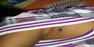
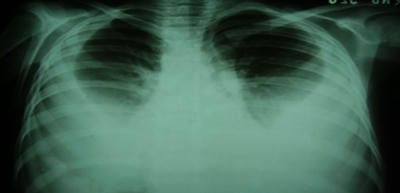
Scrub typhus is caused by Orientia tsutsugamushi and is transmitted to humans by an arthropod vector of the Thrombiculidae family. It is one of the most common re-emerging ricketssial infection in India and other South east Asian countries. Nearly a billion people are at risk with at least a million cases being reported from this region every year. It is distributed in the tsutsugamushi triangle which is distributed over a wide area of 13 million km2. Eschar is the characteristic lesion that starts as a vesicular lesion at the site of mite feeding. It is present in about 40% of cases. It progresses to an ulcer with black necrotic center and an erythematous border along with regional lymphadenopathy. It may affect the central nervous system, cardiovascular system, renal and gastrointestinal system. Here we report three cases which depict the atypical presentations of this disease. It is an eye-opener for clinicians to keep this as a provisional diagnosis in patients who present with fever of unknown origin.

Keywords: Scrub typhus

Corresponding Author How to Cite this Article To Browse
Aroop Mohanty, Senior Resident, Department of Microbiology, All India Institute of Medical Sciences, Rishikesh, Uttarakhand, India.
Email:
Mohanty A, Kabi A, Jha MK, Rekha S, Anusha K R, Gupta P. Scrub typhus – atypical presentations: a case series from the state of Sikkim. Trop J Pathol Microbiol. 2019;5(8):568-573.
Available From
https://pathology.medresearch.in/index.php/jopm/article/view/311

Manuscript Received Review Round 1 Review Round 2 Review Round 3 Accepted
2019-07-28 2019-07-08 2019-07-17 2019-07-21
Conflict of Interest Funding Ethical Approval Plagiarism X-checker Note
No Nil Yes 5%

© 2019 by Aroop Mohanty, Ankita Kabi, Mithilesh Kumar Jha, Sasi Rekha, Anusha K R, Pratima Gupta and Published by Siddharth Health Research and Social Welfare Society. This is an Open Access article licensed under a Creative Commons Attribution 4.0 International License https://creativecommons.org/licenses/by/4.0/ unported [CC BY 4.0].

Introduction

Scrub typhus is a life-threatening zoonosis caused by Orientia tsutsugamushi (formerly Rickettsia) an obligate intracellular Gram-negative bacterium, which was isolated in 1930. It is transmitted to human beings by the bite of a larval Leptotrombidium mite (chigger) [1]. The target cells are the endothelial cells, monocytes and other cell types and the disease is associated with vasculitis and endothelial dysfunction. Illness varies from mild and self-limiting to fatal. After an incubation period of 6-21 days, onset is characterized by fever, rash, headache, myalgia, cough and lymphadenopathy.

Fewer than 50% of patients develop an eschar and less than 40% develop a rash. It is endemic to a geographically distinct region, the so-called tsutsugamushi triangle, which includes Japan, Taiwan, China and South Korea. It also occurs in Nepal, Pakistan, Papua New Guinea, and Australian states. In India, initial reports appeared in the 1930s [2], and a large number of cases were identified among troops during World War II in Assam and West Bengal [3]. Lately it has been reported with increasing frequency from diverse ecologies, initially from southern India and later from the Himalayan belt, the plains of northern India, coastal areas and even from metropolitan cities [4, 5]. The state of Sikkim situated in the Eastern Himalayas has a total geographic area of 7,299 km2 and is essentially a mountainous region without flat piece of land of any extent anywhere.

The mean annual rainfall is maximum in Gangtok (3494mm) and is well distributed during the months of from May to early October. Heavy rainfall along with humid climate in these areas at lower altitude are very conducive to the spread of arthropod vectors.

The first outbreak of Scrub typhus was reported in 2003-2004, when a cluster of pediatric patients presented with fever, hepatospleno-megaly and eschar. Serological confirmation was done in National center of Disease control (NCDC), New Delhi.

Despite a few studies done on this topic from this region, it is still being grossly under-diagnosed due to its non - specific clinical presentation, limited public awareness, and low index of suspicion among clinicians and lack of diagnostic facilities in most places.

We hereby present a case series showing the rare atypical clinical manifestations of Scrub typhus from a tertiary care teaching hospital in Sikkim, India. These manifestations if present in a patient will help the clinicians in implementing a suspect and treat strategy and initiate proper treatment to prevent serious morbidity and fatality in this potentially treatable and curable disease.

Case 1

A 2-year-old male child, presented to the Department of Pediatrics of our Institute with fever associated with chills for 6 days, decreased urine output for 4 days, progressive abdominal swelling associated with loose stools for 3 days and non-productive cough for 2 days and was hospitalized. Physical examination revealed a febrile child with pallor, periorbital puffiness, pedal edema, hepatomegaly, evidence of free fluid in abdomen, and bilateral pleural effusion. Injection Ceftriaxone was started empirically along with supportive treatment, but the child did not improve. Anti-Streptolysin O test was done in order to rule out post streptococcal glomerulonephritis which was negative.

A brownish-black crusted lesion with surrounding erythema-eschar was found below left axilla (Figure-1). Blood samples were sent for routine investigations. Complete blood count revealed an Hb of 9.9gm/dl, TLC of 6,600/mm3, polymorph 85%) and platelets of 90,000/mm3. The urine analysis indicated proteinuria, microscopic hematuria and RBC casts. Blood urea (43mg/dl) and serum creatinine (1.6mg/dl) were raised. The liver function tests revealed transamnitis and cholestasis (ALT 148IU/L, AST 161IU/L). Kidney function also was deranged with sodium (43mg/dl) and potassium (7.8mg/dl).

The Chest X-Ray PA view confirmed bilateral pleural effusion (Figure-2). USG of abdomen and pleural spaces revealed ascites with hepatospleno-megaly and bilateral pleural effusion. Widal test was negative. HbsAg and Anti HCV was negative. A diagnosis of Scrub typhus complicated with AKI was made based on basis 167meqof clinical findings, laboratory results and positive Weil-Felix test and IgM ELISA. Tab Doxycycline was started on 3rd day of admission by 6th day of admission, child became afebrile, with regression of edema and was discharged home on the 10th day.


patho_311_01.jpg

Figure 1: Solitary eschar below left axilla

patho_311_02.jpg

Figure 2: CXR PA view showing B/L pleural effusion

Case 2

A 53-year-old man, presented to the Emergency Department of our Institute with fever, headache, vomiting and altered consciousness since last two days. The informant was his son with fair reliability. The fever was initially low grade but had become high grade and was associated with chills and rigors. He developed confusion and disorientation a day two days before hospitalization. There was no history of seizures, cough or dysuria or rashes. A known hypertensive on regular medications. He was a teetotaler and non-smoker. On examination at admission he was disoriented to time, place and person, was febrile and had BP record of 170/100 mm of Hg. He did not have any icterus, rashes or signs of meningeal irritation. Cranial nerve examination revealed no deficit except bilateral papilledema. Other systemic examination was normal.

Investigations revealed normal Urine examination. CBC revealed TLC of 14,230/cmm polymorph 85%, with normal Hb and platelets. The liver function tests revealed transamnitis and cholestasis (ALT 99U/L, AST 122U/L, GGT 300U/L and ALP of 304U/L). USG of abdomen revealed mild hepatospleno-megaly. Chest X-ray PA view was normal. Leptospira MAT- negative. NCCT of head revealed features of early cerebral edema.

CSF studies revealed glucose 45mg/dL, protein of 78mg/dL, cells of 202/cmm with lymphocytic predominance, negative for AFB, ADA of 5U/L, CBNAT was negative. CSF viral antibodies for HSV, JE were negative. IgM for Scrub on Day 2 of hospitalization was 1.25 (normal range <0.14). He was treated with parenteral Doxycycline, Mannitol and Levetiracetam. He improved slowly and a repeat investigation after one week revealed a normal CSF study, a normal MRI brain and an IgM Scrub titer of 1.68. He improved slowly but completely and was discharged after total two weeks hospitalization.

Case 3

A 45-year-old male who was previously well presented to Emergency department of our Institute with complaints of fever, shortness of breath and cough with scanty expectoration for the last 7 days. The fever was high grade in nature and was occasionally associated with chills and rigors. He was referred from a nearby private nursing home where he was admitted for the last 3 days. During his stay there, he developed aphasia and shortness of breath. There was no history of any seizure, rashes, weakness or hematuria. The patient was a chronic smoker for the past 20 years. On examination he was conscious, alert and oriented. He was febrile and had a heart rate of 120 beats/min, respiratory rate 44 breaths/min and blood pressure 90/60 mm of Hg. The pulse was irregularly irregular. On systemic examination, bilateral coarse crepitation were present in lung bases on auscultation. Other examination were within normal limits. A provisional diagnosis of acute febrile illness was made with an incidental finding of atrial fibrillation (AF). Cardiology opinion was sought for the same.

Patient consent was taken for DC cadioversion and a shock of 200 joules was given. The rate was brought in control, but the AF could not be reverted back. Inj Amiodarone 150 mg was given immediately and then tapered at regular intervals. Blood samples were sent for routine investigations. CBC revealed TLC of 15,400/cmm polymorph 87%, with normal Hb and platelets. The liver function tests were within normal limits. Dengue serology, malarial antigen came negative. Cultures from blood and urine were sterile. Weil-Felix test was positive and IgM antibody for scrub typhus was strongly positive (by ELISA).


The patient was reexamined thoroughly, and an eschar was found on the back of the neck confirming the diagnosis (Figure-3). He was started on doxycycline (100 mg twice a day). He responded to doxycycline and within 48 hours his symptoms decreased significantly and her vitals steadily improved. The patient’s condition improved gradually, and he was discharged after two weeks.

patho_311_03.jpg

Figure 3: Eschar below nape of neck

Discussion

Rickettsial infections are emerging as pathogens around the world in areas which were hitherto unaffected. The rickettsial diseases were once thought to have disappeared from India, but cases are re-emerging from several parts of the country. Scrub typhus is now most commonly reported rickettsial infection from the Indian subcontinent. The diagnosis of scrub typhus is made by using Faine rule, with proper history, epidemiological data, occupational history, clinical examination, seasonal variation and laboratory support. In India, scrub typhus cases have been regularly reported from Vellore and Tamil Nadu [5]. There are scanty reports of scrub typhus from Sikkim state. [6, 7]. Darjeeling is one of the endemic areas of scrub typhus until 1960, thereafter no outbreak was there. In 2005, cases are being reported. Sikkim has annual rainfall of 429-666mm [8]. Vegetation in Sikkim also favors the chiggers to attach with rodents during monsoon season [9]. The outbreaks are associated with the predominance of the vector Leptotrombidium deliense, but recently there has been an important observation L. deliense was missing and there is emergence of Schoengastiella ligula, as the primary vector in the outbreak of Kurseong district of West Bengal [10] Ogawa et al have reported presence of eschars in 7 to 97% of scrub typhus cases in Japan [11]. Multiple eschars have been reported in a single patient [12].

In our case a diligent search for skin rashes helped in finding an eschar and later helped in diagnosis. The majority of studies regarding rickettsial infections from various parts of the world are based on adult populations [13]. There is paucity of studies regarding the incidence and clinical profile of scrub typhus in children of scrub typhus. Scrub typhus is regarded as a life-threatening disease in children.

The complications of scrub typhus are pneumonia, acute respiratory distress syndrome, acute hepatitis, acute kidney injury, meningitis, pancreatitis, acalculous cholecystitis, axonal polyneuropathy, long segment myelitis, DIC, septic shock, MODS [14-17]. In our cases, patients presented with complications with short incubation time. In Indian scenario, there is a higher case fatality of 14% when presenting with complications. In a study conducted in Meghalaya, only 11.1% patients had eschar, 33.3% of patients presented with complications with high mortality [18]. There is need to assess for the predictors of mortality such as blood pressure, live enzymes, platelet count, respiratory effort, serum urea and creatinine in such scenario. The doubling time for scrub typhus is 9-18 hours, so it takes 4 weeks for the culture to become positive [19]. Nested PCR from the eschar samples or buffy coat can help in early diagnosis of the disease within the first 3 days of fever onset even before the appearance of antibodies [20].

Rickettsial infections have been overlooked as a cause of AKI, especially in children. A recent retrospective study from central India did not report any case of AKI in children with Rickettsial infections [21]. Several studies have reported lower incidences of AKI ranging from 2 to 10%. According to Yen H et al. scrub typhus presented with symptoms of acute renal failure is a rare but a serious condition. [22]. The mechanism of AKI in scrub typhus is mainly believed to be impaired renal perfusion due to volume depletion or increased vascular permeability. Overall, renal involvement is considered to be a part of multi-organ dysfunction in patients with severe disease. [23]

Acute myocarditis is associated with scrub typhus more common than previously reported. Patients with high bilirubin and paroxysmal atrial fibrillation (PAF) are at increased risk of acute myocarditis with scrub typhus. And hence these patients should be evaluated for cardiac complications when presented with scrub typhus [24].


The management includes supportive therapy intravenous fluid replacement therapy, mechanical ventilation, and intravenous antibiotics, with addition of Tab. Doxycycline 100mg twice daily for 7 days.

Table-1: Recent studies on scrub typhus

State Year of study No of suspected cases No of lab confirmed cases Eschar Mortality
Rajasthan [25] 2017 NA 66 12% 21.2%
Assam [26] 2017 511 104 0% 49%
Uttar Pradesh [27] 2018 357 97 0% NA
Punjab [28] 2014 772 98 10.2% 3%
Andhra Pradesh [29] 2014-2015 NA   10% NA
Meghalaya [18] 2011-2012 662 90 11.1% 38.5%
Uttarakhand [30] 2015 NA 284 17% 8.5%

Conclusion

Scrub typhus has been under diagnosed in India due to its non-specific clinical presentation, limited awareness and low index of suspicion among clinicians and lack of diagnostic facilities. The clinical manifestations of scrub typhus in children are nonspecific and are likely to be misdiagnosed. Scrub typhus does not find a mention in most descriptions of tropical community acquired AKI or acute myocarditis.

Considering the re-emergence of scrub typhus in India, patients presenting with fever and acute kidney injury as well as rhythm abnormalities should be investigated for scrub typhus. Early diagnosis of scrub typhus and the initiation of empirical therapy with doxycycline will reduce the patient morbidity and mortality.

Declaration of patient consent: The authors certify that they have obtained all appropriate patient consent forms. In the form the patient(s) has/have given his/her consent for his/her/their images and other clinical information to be reported in the journal. The patients understand that their names and initials will not be published and due efforts will be made to conceal their identity, but anonymity cannot be guaranteed.

Reference

01. Weitzel T, Dittrich S, López J, Phuklia W, Martinez-Valdebenito C, Velásquez K, et al. Endemic Scrub Typhus in South America. N Engl J Med. 2016; 375 (10):954-961. doi: 10.1056/NEJMoa1603657 [Crossref][PubMed][Google Scholar]

02. Roy BC. Typhus fever; with special reference to its incidence in India. J Indian Med Assoc. 1945-1946; 15:135-146. [Crossref][PubMed][Google Scholar]

03. Sayen JJ, Pond HS, Forrester JS, Wood FC. Scrub typhus in Assam and Burma: a clinical study of 616 cases. Med. 1946; 25(2):155-214. doi: 10.1097/ 0000 5792 -194605000-00003 [Crossref][PubMed][Google Scholar]

04. Mathai E, Rolain JM, Verghese GM, Abraham OC, Mathai D, Mathai M, et al. Outbreak of scrub typhus in southern India during the cooler months. Ann N Y Acad Sci. 2003;990:359-364. doi:10.1111/j.1749-6632. 2003.tb07391.x [Crossref][PubMed][Google Scholar]

05. Mahajan SK, Rolain JM, Kashyap R, Bakshi D, Sharma V, Prasher BS, et al. Scrub typhus in Himalayas. Emerg Infect Dis. 2006;12(10):1590-1592. doi:10.3201/eid1210.051697 [Crossref][PubMed][Google Scholar]

06. Aggarwal P, Mahesh DM, Ravi Kumar V, Himral P, Kaushal SS, Verma BS. Atypical eschar sites in scrub typhus in sub-Himalayas. J Assoc Physicians India. 2009;57:154. [Crossref][PubMed][Google Scholar]

07. Bhalwar R,Vaidya R, editors. Textbook of public health and community medicine. Department of Com-munity Medicine, Armed Forces Medical College. 2009. [Crossref][PubMed][Google Scholar]

08. Mean monthly meteorological parameters of gangtok period:1971-2000. Available from: http://imdsikkim. gov. in/monthlymetparam. Html [Last accessed on 2012 Oct 16] [Crossref][PubMed][Google Scholar]

09. Frances SP, Watcharapichat P, Phulsuksombati D, Tanskul P, Linthicum KJ. Seasonal occurrence of Leptotrombidium deliense (Acari: Trombiculidae) attached to sentinel rodents in an orchard near Bangkok, Thailand. J Med Entomol. 1999;36(6):869-874. doi: 10.1093/jmedent/ 36.6.869 [Crossref][PubMed][Google Scholar]


10. Rina Tilak, Rajesh Kunwar, Urmila B Wankhade, V W Tilak. Emergence of Schoengastiella ligula as the vector of scrub typhus outbreak in Darjeeling: has Leptotrom-bidium deliense been replaced?. Indian J Public Health. 2011;55(2):92-99. doi: 10.4103/0019-557X.85239 [Crossref][PubMed][Google Scholar]

11. Mahajan SK. Scrub typhus. J Assoc Physicians India. 2005;53:954-958. [Crossref][PubMed][Google Scholar]

12. Gurung S, Pradhan J, Bhutia PY. Outbreak of scrub typhus in the North East Himalayan region-Sikkim: an emerging threat. Indian J Med Microbiol. 2013;31 (1): 72-74. doi: 10.4103/0255-0857.108729 [Crossref][PubMed][Google Scholar]

13. Sayen Jj, Pond HS. Scrub typhus in Assam and Burma; a clinical study of 616 cases. Medicine (Baltimore). 1946;25:155-214. doi:10.1097/00005792-194605000-00003 [Crossref][PubMed][Google Scholar]

14. Dhakal M, Dhakal OP, Bhandari D. Pancreatitis in scrub typhus: a rare complication. BMJ Case Rep. 2014; 2014. pii: bcr2013201849. doi: 10.1136/bcr-2013-201849 [Crossref][PubMed][Google Scholar]

15. Tandon R, Kumar A, Kumar A. Long-Segment Myelitis, Meningoencephalitis, and Axonal Polyneuro-pathy in a Case of Scrub Typhus. Ann Indian Acad Neurol. 2019;22(2):237-240. doi: 10.4103/aian. AIAN_ 66_18 [Crossref][PubMed][Google Scholar]

16. Chi WC, Huang JJ, Sung JM, Lan RR, Ko WC, Chen FF. Scrub typhus associated with multiorgan failure: a case report. Scand J Infect Dis. 1997;29 (6):634-635. doi:10.3109/00365549709035911 [Crossref][PubMed][Google Scholar]

17. Yi SY, Tae JH. Pancreatic abscess following scrub typhus associated with multiorgan failure. World J Gastroenterol: WJG. 2007;13(25):3523-3525. doi: 10. 3748/ wjg.v 13.i25.3523 [Crossref][PubMed][Google Scholar]

18. Sivarajan S, Shivalli S, Bhuyan D, Mawlong M, Barman R. Clinical and paraclinical profile, and predictors of outcome in 90 cases of scrub typhus, Meghalaya, India. Infect Dis Poverty. 2016;5(1):91. doi: 10.1186/s40249-016-0186-x [Crossref][PubMed][Google Scholar]

19. Tamura A, Ohashi N, Urakami H, Miyamura S. Classification of Rickettsia tsutsugamushi in a new genus, Orientia gen nov, as Orientia tsutsugamushi comb nov. Int J Syst Bacteriol. 1995;45(3):589-591. doi: 10.1099/00207713-45-3-589 [Crossref][PubMed][Google Scholar]

20. Kim DM, Kim SW, Choi SH, Yun NR. Clinical and laboratory findings associated with severe scrub typhus. BMC Infect Dis. 2010;10:108. doi: 10.1186/1471-2334-10-108 [Crossref][PubMed][Google Scholar]

21. Rathi NB, Rathi AN, Goodman MH, Aghai ZH. Rickettsial diseases in central India: proposed clinical scoring system for early detection of spotted fever. Indian Pediatr. 2011;48(11):867-872. [Crossref][PubMed][Google Scholar]

22. Yen TH, Chang CT, Lin JL, Jiang JR, Lee KF. Scrub typhus: a frequently overlooked cause of acute renal failure. Ren Fail. 2003;25(3):397-410. doi: 10.1081/jdi-120021152 [Crossref][PubMed][Google Scholar]

23. Mathai E, Rolain JM, Verghese GM, Abraham OC, Mathai D, Mathai M, et al. Outbreak of scrub typhus in southern India during the cooler months. Ann N Y Acad Sci. 2003;990(1):359-364. [Crossref][PubMed][Google Scholar]

24. Jung YeonChin,Ki-Woon Kang, Kyung Min Moon, Jongwoo Kim, Yu Jeong Choi. Predictors of acute myocarditis in complicated scrub typhus: an endemic province in the Republic of Korea. Korean J Intern Med. 2018;33(2):323–330. doi: 10.3904/ kjim. 2016.303 [Crossref][PubMed][Google Scholar]

25. Takhar RP, Bunkar ML, Arya S, Mirdha N, Mohd A. Scrub typhus: A prospective, observational study during an outbreak in Rajasthan, India. Natl Med J India. 2017;30(2):69-72. [Crossref][PubMed][Google Scholar]

26. Khan SA, Bora T, Laskar B, Khan AM, Dutta P. Scrub Typhus Leading to Acute Encephalitis Syndrome, Assam, India. Emerg Infect Dis. 2017;23(1):148-150. doi: 10.3201/eid2301.161 038. [Crossref][PubMed][Google Scholar]

27. Rizvi M, Sultan A, Chowdhry M, Azam M, Khan F, Shukla I, et al. Prevalence of scrub typhus in pyrexia of unknown origin and assessment of interleukin-8, tumor necrosis factor-alpha, and interferon-gamma levels in scrub typhus-positive patients. Indian J Pathol Microbiol. 2018;61(1):76-80. doi: 10.4103/IJPM.IJPM_644_16 [Crossref][PubMed][Google Scholar]


28. Oberoi A, Varghese SR. Scrub typhus-an emerging entity: a study from a tertiary care hospital in North India. Indian J Public Health. 2014;58(4):281-283. doi: 10.4103/0019-557X.146299 [Crossref][PubMed][Google Scholar]

29. Peesapati N, Lakkapraga R, Sunitha S, Sivaram PV. Clinical manifestations and complications of scrub typhus- A hospital-based study from North Andhra. Astrocyte. 2015;2(3):116-120. doi: 10.4103/2349-0977. 181508 [Crossref][PubMed][Google Scholar]

30. Bhargava A, Kaushik R, Kaushik RM, Sharma A, Ahmad S, Dhar M, Mittal G, Khanduri S, Pant P, Kakkar R. Scrub typhus in Uttarakhand & adjoining Uttar Pradesh: Seasonality, clinical presentations & predictors of mortality. The Indian J Med Res. 2016; 144(6):901-909. doi: 10.4103/ijmr.IJMR_1764_15 [Crossref][PubMed][Google Scholar]