E-ISSN:2456-1487
P-ISSN:2456-9887
RNI:MPENG/2017/70771

Research Article

Tumor

Tropical Journal of Pathology and Microbiology

2019 Volume 5 Number 8 August
Publisherwww.medresearch.in

Small round cell tumor of cervical region: a case report with unusual site & course

D. Pathak V.1, Metkar G.2*, Bhide S.3, Joshi S.4
DOI: https://doi.org/10.17511/jopm.2019.i08.09

1 Vikas D. Pathak, Department of Pathology, MIMER Medical College, Pune, Maharashtra, India.

2* Gauri Metkar, Department of Pathology, MIMER Medical College, Pune, Maharashtra, India.

3 Smita Bhide, Department of Pathology, MIMER Medical College, Pune, Maharashtra, India.

4 Sneha Joshi, Department of Pathology, MIMER Medical College, Pune, Maharashtra, India.

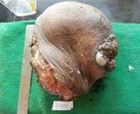
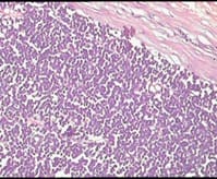
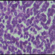
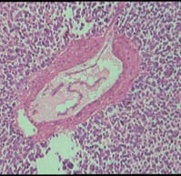
Ewing’s sarcoma belongs to a spectrum of neoplastic diseases known as Ewing’s family of tumors. Ewing sarcoma can arise in either bone or soft tissue. Small round cell tumors comprise a heterogeneous group of neoplasms that predominate in childhood and adolescence, share similar morphological features, consisting of dense cellular proliferation of small round cells with primitive appearance. Ewing sarcoma is the second most common malignant bone tumour in children and young adults, although, rarely, it may be of extraskeletal origin. Extra-osseous tumor with metastatic or recurrent disease has a worse outcome; 5-year overall survival remains about 25%. A female patient presented in surgical OPD for the first time at the age of 12 years with swelling in right supraclavicular region. The swelling was excised & a provisional diagnosis of small round cell tumor was given & patient was lost to follow up. Swelling recurred at the age of 23 years & was gradually increasing in size, measuring 20 x 20 x 20 cm. Radiologically, lesion was suggestive of sarcoma / lymphoma. Patient was operated & histopathological diagnosis of malignant round cell tumor was given with differential diagnosis of Ewing’s sarcoma, PNET, lymphoma. Patient had third recurrence of swelling at the age of 28 years at same site. On IHC, diagnosis of Soft Tissue Ewing sarcoma was given. Inspite of recurrence & huge size of tumor, patient was otherwise asymptomatic. Ours is a rare case of more than 16 years of survival inspite of recurrence.

Keywords: Small round cell tumor, Ewing sarcoma, Extra-osseous tumor, Immunohistochemistry

Corresponding Author How to Cite this Article To Browse
Gauri Metkar, Department of Pathology, MIMER Medical College, Pune, Maharashtra, India.
Email:
Pathak VD, Metkar G, Bhide S, Joshi S. Small round cell tumor of cervical region: a case report with unusual site & course. Trop J Pathol Microbiol. 2019;5(8):562-567.
Available From
https://pathology.medresearch.in/index.php/jopm/article/view/310

Manuscript Received Review Round 1 Review Round 2 Review Round 3 Accepted
2019-08-05 2019-08-15 2019-08-24 2019-08-27
Conflict of Interest Funding Ethical Approval Plagiarism X-checker Note
No Nil Yes 8%

© 2019 by Vikas D. Pathak, Gauri Metkar, Smita Bhide, Sneha Joshi and Published by Siddharth Health Research and Social Welfare Society. This is an Open Access article licensed under a Creative Commons Attribution 4.0 International License https://creativecommons.org/licenses/by/4.0/ unported [CC BY 4.0].

Introduction

Ewing sarcoma was first described in 1921 by James Ewing. They arise from mesenchymal progenitor cells and are part of a spectrum of neoplastic diseases, known as Ewing’s sarcoma family of tumors (ESFT) [1]. Ewing's sarcoma family of tumors (ESFT) represents a family of morphologically similar small round-cell neoplasms, including classic Ewing's sarcoma (ES) of the bone next to extraskeletal ES, small-cell tumor of the thoracopulmonary region (Askin tumor) and soft-tissue-based primitive neuroectodermal tumors (PNETs). ESFT are characterized by reciprocal translocation between chromosome 11 & 22, t (11; 22), and are usually seen at osseous sites, both axial and appendicular. Extra- osseous presentation is uncommon.

Ewing sarcoma is the second most common childhood primary bone cancer, though a substantial proportion of these tumors arise from extraskeletal sites [2]. Ewing sarcoma usually arises in bone and is associated with soft tissue extension in 90% of cases. EES arises in the soft tissues of the trunk or extremities. Although it is a soft tissue primary tumor, it can cause changes in the cortex of the adjacent bone [3-5]. The most common sites are chest wall, paravertebral region, retroperitoneal space, lower extremities, and gluteal region. However, few cases have been reported in the kidney, breast, gastrointestinal tract, prostate, endometrium, the adrenal glands, brain, and lungs [6]. The head and neck region is an unusual primary site for this type of tumor.

ES also occurs as a primary soft tissue neoplasm without involvement of bone [7]. Ewing sarcoma (ES) is a poorly differentiated, highly malignant, round cell tumor without cellular or structural differentiation [8]. Histological assessment shows that these (EES) tumors are composed of sheets of small round cells with hyperchromatic nuclei, and scanty cytoplasm.

Immunohistochemically, a number of markers have been used, of which MIC2 (CD99) is particularly useful in eliciting membranous positive staining of tumor cells [9]. Here, authors present a case of extraskeletal Ewing sarcoma presenting in right supraclavicular region in a 23 years female with recurrence of same tumor at same site without bone involvement or metastases from any other place with 16 years of survival.

Case Report

History: A female patient presented in surgical OPD with swelling in the right supraclavicular region, for the first time at age of 12 years. Local excision of mass was done & diagnosis of small round cell tumor was given & patient was advised IHC analysis. Subsequently patient was lost to follow up. Swelling recurred and patient again came to OPD after 11 years at the age of 23 years with swelling gradually increasing in size measuring 23 x 17 x 11 cm at time of presentation. All routine investigations were within normal limits.

CT scan of Neck region: 13 x 18 x 11 cm size exophytic well encapsulated solid cystic lesion in right supraclavicular region with impression S/O lymphoma.

MRI Neck & Chest: tumor was seen in subcutaneous & intramuscular planes of neck and chest wall suggestive of sarcoma. Bone was not involved.

Treatment: Wide local excision of swelling was done.

Gross Features: Excised swelling was received in our Department of Pathology measuring 23 x 16.5 x 11 cm, external surface partially encapsulated and nodular (Figure 1). Cut surface hemorrhagic with greyish white soft to firm areas with tumor deposits outside the capsule & involving the base.

patho_310_01.jpg

Figure-1: Photograph showing external surface partially encapsulated and nodular

Microscopic Features: Tumor was partially encapsulated and composed of sheets of monomorphic, round cells with large hyperchromatic nuclei and scanty cytoplasm (Figure 2, 3).


Pseudorosette formation was noted in the tumor (Fig. 4). Mitotic count was 1-2/10 HPF and necrosis was not seen. Lymphovascular invasion not seen. Base was involved by tumor.

patho_310_02.jpg

Figure 2: Photomicrograph showing partially encapsulated tumor mass and composed of sheets of monomorphic, round cells with large hyperchromatic nuclei and scanty cytoplasm. (H&E, 5X).

patho_310_03.jpg

Figure 3: Photomicrograph showing tumor mass composed of sheets of monomorphic, round cells with large hyperchromatic nuclei and scanty cytoplasm. (H&E, 40X)

Figure 4: Photomicrograph showing Pseudorosette formation. (H&E, 10X)

patho_310_04.jpg

Special Stain: PAS Stain- Positive.

TNM classification

· pT2b - Tumour size more than 5cm in greatest dimensions.

· pNx - Regional Lymph Node

· pMx- Distant metastasis

· Histological grading: Grade II

· Stage Grouping: Stage IIB.

Impression: Malignant small round cell tumor.

Differential Diagnosis

  • Ewing sarcoma
  • PNET
  • Lymphoma.

Advice: Immunohistochemistry (IHC) such as MIC2, Cytokeratin, CD20, CD3, Synaptophysin, Desmin for final diagnosis.

The tumor was examined for IHC marker with following results:

Positive: MIC2 (diffusely) & Cytokeratin (weakly & focally)

Negative: CD20, CD3, Synaptophysin, Desmin.

Impression on Immunohistochemistry (IHC): Soft Tissue Ewing Sarcoma.

Final Diagnosis: Soft tissue (Extraskeletal) Ewing sarcoma.


Discussion

The diagnosis and treatment of soft tissue small cell sarcomas continues to be a challenge to the pathologist and the medical and surgical oncologist. Malignant small round cell tumor are characterized by small, round, relatively undifferentiated cells. They include Ewing’s sarcoma, peripheral neuroectodermal tumor, rhabdomyosarcoma, synovial sarcoma, Non–Hodgkin’s lymphoma, neuroblastoma, hepatoblastoma and nephroblastoma.

On histopathological examination these tumors appear similar due to morphological similarities. Thus, for further typing of tumor one should do special stains, immunohistochemistry, fluorescence in situ hybri-dization or electron microscopic examination. Once the specific diagnosis is made then appropriate treatment and management can be approached. It also helps to assess the prognosis of the patient.

Allam et al has reported a male to female ratio of 2.4:1 in a series of 24 patients [10].

Approximately 80% of patients are diagnosed with Ewing sarcoma at a younger age of less than 20 years. A total of 50-60% patients are diagnosed during their second decades [11].

Tefft et al first described EES in 1969 to be histologically similar to primary Ewing’s sarcoma of bone [3].

Here a female patient who was diagnosed at the age of 12 years with small round cell tumor. Radiological investigation like MRI was also suggestive of sarcoma with no bone involvement. MRI is the most sensitive test available for the evaluation of the soft tissue extent of the tumor [12].

In this present case, tumor recurred after 16 years at the same site without any complication. Surgical removal was done as it is the treatment of choice [13] for EES, followed by adjunctive chemotherapy and radiotherapy.

Around 75% patients present with rapidly growing painless mass, 30% patients exhibiting distant metastasis at the time of diagnosis. Clinically, patients with EES usually present with a painless, rapidly growing mass but tumors arising elsewhere may be painful.

Our patient presented with a slow growing mass in the right side of neck.

EES is a tumor sensitive to multimodality treatment. Early awareness and wide

resection followed by chemotherapy and radiotherapy might improve the long-term survival of patients with extraskeletal Ewing’s sarcoma. Although the prognosis for this tumor is poor, an early and adequate surgical resection followed by adjunctive chemotherapy and radiotherapy for microscopically positive surgical margins improve the survival rate [3, 14].

Relapse rate is 30% after treatment, considering that most of the relapsed patients do not survive. Patients with metastatic or recurrent disease have a worse outcome. The overall 5-years survival rate remains about 25%. [15].

EES is a rare soft tissue neoplasm that can develop in the soft tissues at any location. Its occurrence in head and neck as a primary tumor is very unusual. Distant metastases are also common in EES.

In our patient tumor was located in right supraclavicular region. The head and neck region is an unusual primary site for this type of tumor and, according to Chao et al, there are only five out of 118 cases of EES located in head and neck region [16].

Moreover, primary cervical localization is considered extremely rare, with only 13 cases reported in medical literature from 1969 to 2015 [17]. Only one case has been reported in parapharyngeal space [18].

Rud et al. in a large series of 42 cases of EES reported few cases in head and neck region [19].

Haytham Eloqayli in 2017 has reported EES in cervical region [20].

Nazia Mashriqi et al in 2015 observed that on follow-up of these (EES) cases ranging between 5 months and 8 years, with 15 of the 19 cases being alive and recurrence-free at the time of follow-up.

Two patients died of metastatic disease 12 days and 4.2 years after presentation, respectively. Patient developed metastatic disease while on adjuvant chemo-therapy and died 10 months after diagnosis [21].

Rud et al in 1989 observed that in group 2 one patient treated by local resection, irradiation, and chemotherapy is alive without disease 7.8 years after surgery.


The remaining two patients died within 6 months of surgery. One patient died from brain metastases 5 months after surgery and one patient died from treatment toxicity [19].

In this case diagnosis of Ewing sarcoma was made on immunohistochemical staining as described above. Inspite of recurrence & huge size of tumor, patient was otherwise asymptomatic. As Ewing sarcoma has an aggressive course especially after recurrence, ours is a rare case of more than 16 years survival inspite of recurrence without any complication.

Conclusion

Primary cervical EES is rare. No specific treatment guidelines have been established for EES. Consequently, treatment depends on patient’s age, progression of tumor & its complications. Multimo-dality such as X-ray, MRI, CT scan and FNAC were used for diagnosis & the definitive diagnosis was made on histopathology & IHC (Immunohistochemistry) examination. IHC is mandatory for diagnosis of soft tissue Ewing’s sarcoma in younger age group patients. Regular follow up of such patients is essential for timely detection & complete treatment of recurrent tumors. The aim should be to improve or preserve the quality of life and long-term survival of patients.

Contribution by different authors

Dr. Vikas D. Pathak and Dr. Gauri Metkar: Histopathology reporting, follow up of case and manuscript preparation. Dr. Smita Bhide and Dr. Sneha Joshi: Guidance in manuscript preparation.

Reference

01. Jedlicka P. Ewing Sarcoma, an enigmatic malignancy of likely progenitor cell origin, driven by transcription factor oncogenic fusions. Int J Clinic Experiment Pathol. 2010;3(4):338-347. [Crossref][PubMed][Google Scholar]

02. Granowetter L, Womer R, Devidas M, Krailo M, Wang C, Bernstein M, et al. Dose-intensified compared with standard chemotherapy for nonmetastatic Ewing sarcoma family of tumors: a Children's Oncology Group Study. J Clinic Oncol. 2009;27(15):2536-2541. doi: 10.1200/JCO.2008.19.1478 [Crossref][PubMed][Google Scholar]

03. Tefft M, Vawter GF, Mitus A. Paravertebral "round cell" tumors in children. Radiol. 1969;92(7):1501-1509. doi: 10.1148/92.7.1501 [Crossref][PubMed][Google Scholar]

04. Angervall L, Enzinger FM. Extraskeletal neoplasm resembling Ewing's sarcoma. Cancer. 1975;36(1):240-251. doi: 10.1002/1097-0142(197507)36:1<240::aid-cncr2820360127>3.0.co;2-h [Crossref][PubMed][Google Scholar]

05. Soule EH, Newton Jr W, Moon TE, Tefft M. Extraskeletal Ewing's sarcoma- A preliminary review of 26 cases encountered in the inter group rhabdomyo-sarcoma study. Cancer. 1978;42(1):259-264. doi: 10.3892/ol.2015.4013 [Crossref][PubMed][Google Scholar]

06. Basma EE, Hajar H, Nabil M, Anis B, Samir B. Breast Ewing Sarcoma / Primitive Neuroectodermal Tumor: A Case Report and a Review of the Literature. Breast. 2012;3(10):WMC003792. [Crossref][PubMed][Google Scholar]

07. Geens L, Robays JV, Geert V, der Speeten KV. An Unusual Location of Extraosseous Ewing's Sarcoma. CaseRepOncol. 2013;6(2):293-302. doi: 10.1159/000351836 [Crossref][PubMed][Google Scholar]

08. Paoletti H, Colineau X, Acalet L, Tourrette JH, Civatte M, Fesselet J, et al. [Ewing's sarcoma of the soft tissues: apropos of 3 cases and review of the literature]. J Radiol. 1999;80(5):477-482. [Crossref][PubMed][Google Scholar]

09. Rao TD, Padma M. Abdominal Wall Extraskeletal Ewing Sarcoma - Case Report. IOSR J Dent Med Sci (IOSR-JDMS). 2015;14(1):4-6. [Crossref][PubMed][Google Scholar]

10. AllamA, El-Husseiny G, Khafaga Y, Kandil A, Gray A, Ezzat A, et al. Ewing's Sarcoma of the Head and Neck: A Retrospective Analysis of 24 Cases. Sarcoma. 1999; 3(1):11-15. doi: 10.1080/13577149977811 [Crossref][PubMed][Google Scholar]

11. Iwamoto Y. Diagnosis and treatment of Ewing's sarcoma. Japan J Clin Oncol. 2007;37(2):79-89. doi: 10.1093/jjco/hyl142 [Crossref][PubMed][Google Scholar]

12. Fleming DJ, Murphy MD, Nallu S, Niscastri AD. Primary Ewing sarcoma of lumbar spine with massive intraspinal extension. Pediatr Neurol. 2008;38:58-60. [Crossref][PubMed][Google Scholar]


13. Mattogno PP, Nasi D, Iaccarino C, Oretti G, Santoro L, Romano A. First Case of Primary Sellar/ Suprasellar-Intraventricular Ewing Sarcoma: Case Report and Review of the Literature. World Neurosurg. 2017; 98:869. e1-869.e5. doi: 10.1016/j.wneu.2016.12.045 [Crossref][PubMed][Google Scholar]

14. Raney RB, Asmar L, Newton WA Jr, Bagwell C, Breneman JC, Crist W, et al. Ewing's sarcoma of soft tissues in childhood: a report from the Intergroup Rhabdomyosarcoma Study, 1972 to 1991. J Clin Oncol. 1997; 15(2): 574-582. doi: 10.1200/JCO.1997.15.2. 574 [Crossref][PubMed][Google Scholar]

15. Lee J, Hoang BH, Ziogas A, Zell JA. Analysis of prognostic factors in Ewing sarcoma using a population-based cancer registry. Cancer. 2010;116(8): 1964-1973. doi: 10.1002/cncr.24937 [Crossref][PubMed][Google Scholar]

16. Cho SI, Park YH, Cho JH, Ryoo BY, Yang SH, Youn SM et al. Extraskeletal Ewing’s Sarcoma of the Head and Neck Presenting as Blindness. Korean J Int Med. 2007;22(2):133-137. doi: 10.3904/kjim.2007.22.2.133 [Crossref][PubMed][Google Scholar]

17. Bustoros M, Thomas C, Frenster J, Modrek AS, Bayin NS, Snuderl M et al. Adult primary spinal epidural extraosseous Ewing’s sarcoma: a case report and review of the literature, Case Rep. Neurol. Med. 2016;8. doi: [Article][Crossref][PubMed][Google Scholar]

18. Ng SH, Ko SF, Cheung YC, Wong HF, Jung SM. Extraskeletal Ewing's sarcoma of the parapharyngeal space. Br J Radiol. 2004;77(924):1046-1049. doi: 10.1259/bjr/16676268 [Crossref][PubMed][Google Scholar]

19. Rud NP, Reiman HM, Pritchard DJ, Frassica FJ, Smithson WA. Extraosseous Ewing's sarcoma- A study of 42 cases. Cancer. 1989;64(7):1548-53. doi: 10.1002/1097-0142(19891001)64:7<1548::aid-cncr2820640733>3.0.co;2-w [Crossref][PubMed][Google Scholar]

20. Eloqayli H. Adult primary cervical extra-osseous Ewing’s sarcoma: A case report and short literature review. Int J Surgery Case Reports. 2017;41:83-85. doi: 10.1016/j.ijscr.2017.09.033 [Crossref][PubMed][Google Scholar]

21. Mashriqi N, Gujjarlapudi JK, Sidhu J, Zur M, Yalamanchili M. Ewing’s sarcoma of the cervix, a diagnostic dilemma: a case report and review of the literature. J Med Case Rep. 2015;9:255. doi: 10.1186/s13256-015-0733-2 [Crossref][PubMed][Google Scholar]