E-ISSN:2456-1487
P-ISSN:2456-9887
RNI:MPENG/2017/70771

Research Article

Uterine

Tropical Journal of Pathology and Microbiology

2019 Volume 5 Number 8 August
Publisherwww.medresearch.in

Uterine leiomyomas: a demographical and clinico-pathological exploration in a rustic setup of Tamil Nadu: a comprehensive study

Sankar R.1, Kulandaivel A.2, Krishnaswamy B.3*, Viswanathan P.4
DOI: https://doi.org/10.17511/jopm.2019.i08.15

1 R. Gowri Sankar, Assistant Professor, Department of Pathology, Vinayaka Missions Kirupananda Variyar Medical College and Hospital, Salem, Tamil Nadu, India.

2 Anbu Lenin Kulandaivel, Associate Professor, Department of Pathology, Vinayaka Missions Kirupananda Variyar Medical College and Hospital, Salem, Tamil Nadu, India.

3* B. Krishnaswamy, Professor and Head, Department of Pathology, Rajah Muthiah Medical College and Hospital, Chidambaram, Tamil Nadu, India.

4 P. Viswanathan, Retired Professor, Department of Pathology, Rajah Muthiah Medical College and Hospital, Chidambaram, Tamil Nadu, India.

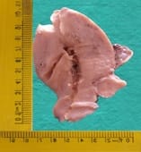
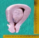
Aim: The aim of this study is to evaluate the prevalence of uterine leiomyomas in a rural setup of Tamil Nadu and also to analyse its plethora among the various neoplastic lesions of female genital tract as well as the various demographical and clinico-pathological spectrum of the same. Material and Methods: A twenty-five years retrospective study was conducted in the Department of Pathology in a rural teaching institute, Rajah Muthiah Medical College and Hospital, in Chidambaram. Result: 1200 cases were studied in the study period among the total two thousand and twenty-two female genital tract neoplastic lesions that were received in the department during the twenty-five-year period. The most common age group affected was 5th decade. The most common clinical presentation was menstrual disturbances among which menorrhagia was the commonest symptom. Taking into account the number of leiomyomas present in a uterus, single leiomyomas outnumbered multiple ones by a vast margin and taking the anatomical location of leiomyoma in a uterus into consideration, intramural leiomyoma stood well ahead of submucosal and subserosal leiomyomas. Conclusion: Almost 2/3rd of the total female genital tract neoplastic lesions received from the inception of college to December 2012 were Uterine leiomyomas there by making it the most common neoplasm of female genital tract reported in our hospital in a rural setup for a total period of twenty five years starting from the inception of college until December 2012.

Keywords: Intramural, Leiomyoma, Menorrhagia, Voluminous study, Rural Tamil Nadu

Corresponding Author How to Cite this Article To Browse
B. Krishnaswamy, Professor and Head, Department of Pathology, Rajah Muthiah Medical College and Hospital, Chidambaram, Tamil Nadu, India.
Email:
Sankar RG, Kulandaivel AL, Krishnaswamy B, Viswanathan P. Uterine leiomyomas: a demographical and clinico-pathological exploration in a rustic setup of Tamil Nadu: a comprehensive study. Trop J Pathol Microbiol. 2019;5(8):600-606.
Available From https://pathology.medresearch. in/index.php/jopm/article/view/309

Manuscript Received Review Round 1 Review Round 2 Review Round 3 Accepted
2019-08-05 2019-08-14 2019-08-20 2019-08-26
Conflict of Interest Funding Ethical Approval Plagiarism X-checker Note
No Nil Yes 8%

© 2019 by R. Gowri Sankar, Anbu Lenin Kulandaivel, B. Krishnaswamy, P. Viswanathan and Published by Siddharth Health Research and Social Welfare Society. This is an Open Access article licensed under a Creative Commons Attribution 4.0 International License https://creativecommons.org/licenses/by/4.0/ unported [CC BY 4.0].

Introduction

Benign tumour of the muscular uterine wall is known by various names such as Leiomyomata/ myxoma/ fibroid [1]. Most of them are detected in middle aged women and are uncommon in women less than thirty years of age [2]. The overall incidence is between 4% and 11%, but it rises to nearly 40% in women over the age of fifty years. Clinically apparent lesions are less common in parous and premenstrual women [3].

The symptomatology may vary based on the location mainly [2]. It ranges from asymptomatic to various menstrual disturbances and even urinary difficulties. They are known to shrink after menopause and it is associated with fibrosis and reduction in individual tumour cells [4, 5]. The frequency does not decrease after menopause [2]. The normal myometrium of leiomyoma containing uterus expresses high level of oestrogen receptors, a fact that may be related to their pathogenesis [6]. The ovarian follicular hormone acts upon myometrium and results in cellular metaplasia with subsequent development of uterine fibroids [7].

Symptomatic leiomyomas need urgent attention by myomectomy in younger females whereas hysterectomy still remains the traditional modality of treatment [8, 9]. The intent of this voluminous study is to estimate the prevalence of uterine leiomyomas, among the hysterectomy specimens received in a rural population setup in the State of Tamil Nadu. This study also analyses the leiomyomas in context to various individual characteristics like age, location, number, clinical presentation as well as some combined parameters that include location and number, location and clinical presentation. To the best of the author’s apprehension such an extensive study involving a rural population in the state have not been done previously. Hence it is being presented as an account of its rarity.

Material and Methods

Study place: The study is conducted in Department of Pathology, Rajah Muthiah Medical College and Hospital, Chidambaram.

Study type: This is a retrospective study.

Study duration: The study duration was for twenty-five years from January 1988 to December 2012.

Inclusion criteria: A total of 1200 hysterectomy specimens diagnosed clinically as uterine leiomyomas that were received in Department of Pathology.

Exclusion criteria: Hysterectomy specimens with other female genital tract conditions were excluded.

Study conduct: The study was a retrospective study from January 1988 to December 2012. The relevant clinical data were retrieved from histopathological requisition forms and clinical records.

Sample size: A total of 1200 hysterectomy specimens received in the Department of Pathology, Rajah Muthiah Medical College and Hospital were included.

All the specimens were fixed in 10% formalin and a detailed examination by two independent Pathologists was done with reference to number, location and combination of fibroids.

Statistical analysis: Data’s were collected and analysed.

Results

A total of 1200 histopathologically proven leiomyoma cases were studied. In the above said period, a total of 2022 neoplastic lesions of female genital tract were received in the Department of Pathology, RMMCH. Majority were benign lesions (1419 cases, 70.2%) (Table-1). Leiomyoma was the most common benign tumor accounting for 84.6% among the benign lesions (Table-2) and 59.3% among all the neoplastic lesions.

Table-1: Total benign vs malignant lesions

Total number of female genital tract neoplastic lesions Benign lesions Malignant lesions
2022 1419 (70.2%) 603 (29.8%)

Table-2: Uterine Leiomyoma vs Other benign lesions

Total number of benign lesions Uterine leiomyomas Other benign lesions
1419 1200 (84.6%) 219 (15.4%)

The age group of the patients were divided into five groups and the highest incidence was found in the 5th decade followed closely by 4th decade (Table-3). The youngest and oldest patient was 19 and 75 years respectively.


In the present study 61% of the patients presented with menstrual disturbances with menorrhagia being the most common symptom.

Table-3: Leiomyomas: age distribution

Age in years Total no. of cases Percentage %
20 & below 01 0.08
21-30 84 7.00
31-40 518 43.20
41-50 530 44.20
51 & above 67 5.52

In the present study, majority were intramural leiomyoma (73.07%) followed by subserosal (14.75%) and submucosal (12.16) leiomyoma (Table-4). The five-year interval study showed a gradual decline in intramural leiomyoma while submucosal and subserosal were slightly increasing as the years passed by (Table-5).

Table-4: Fibroids - location

S. No Type of leiomyoma Total no. of cases Percentage %
1 Intramural 877 73.09
2 Submucosal 146 12.16
3 Subserosal 177 14.75

Table-5: Fibroids - Location (5 Year Interval)

5-year period Intramural Submucosal Subserosal
Total no. of cases % Total no. of cases % Total no. of cases %
1987-1992 88 81.4 08 7.4 12 11.2
1993-1997 103 75.2 12 8.8 22 16
1998-2002 211 72.2 38 13.0 43 14.8
2003-2007 157 74.4 26 12.3 28 13.3
2008-2012 318 70.4 62 13.7 72 15.9

The present study showed 902 cases having only one leiomyoma while 298 cases were found to have more than one leiomyoma among which majority were within single anatomical location while some were seen in combination with other anatomical locations (Table-6). The most common combination seen was intramural with subserosal followed by intramural with submucosal. The number of leiomyomas in context to the location was shown in Table-7.

Table-6: Leiomyomas – solitary vs multiple

S. No Number of fibroids Total no. of cases Percentage %
1 Single 902 75.20
2 Multiple 298 24.80

Table-7: Leiomyomas-location and number

  Single Multiple
Total no. of cases % Total no. of cases %
Intramural 633 72.2 244 27.8
Submucosal 127 87.0 19 13.0
Subserosal 142 80.2 35 19.8

Gross Images

patho_309_01.jpg

Figure-1: Intramural leiomyoma

patho_309_02.jpg

Figure-2: Subserosal leiomyoma


patho_309_03.jpg

Figure-3: Submucosal leiomyoma

patho_309_04.jpg

Figure-4: Intramural and Subserosal

Discussion

Hysterectomy, one of the major gynaecological surgery, was first performed in the year 1843 in England by Charles Clay [10]. Inspite of the availability of medical and conservative management, hysterectomy remains the second most frequently performed obstetric surgery after caesarean section in many parts of the world [11]. The commonest indication for hysterectomy is Leiomyoma [12]. Leiomyoma is a benign neoplasm composed of smooth muscle cells in a variable amount of fibrous stroma [13].

Leiomyomas are encountered frequently in multiparous women between third and fourth decades of life [14]. The National Institute of Environmental Health Sciences (NIEHS) uterine fibroid study provided data on the cumulative incidence of fibroids and documented that black women are more likely than white women to have uterine fibroids [15]. Inspite of Uterine Leiomyomas being highly prevalent and also having an enormous impact on the healthcare system and the economy, there is no effective medical treatment available to eliminate fibroids. This is due in part to the fact that very little is known about their pathophysiology [16]. Studies using glucose-6-phosphate dehydrogenase isoenzyme suggest that each apparently arise from a single cell within the myometrium [17]. Recent studies have confirmed that the different cell types contained in fibroids are all clonally derived from a parental cell with implied multipotent stem cell properties [18]. Although the initial event is believed to be smooth muscle cell proliferation, it is thought that a complex signalling system is also necessary. Thus, there are at least two components in the development of fibroids: the transformation of normal myocytes into abnormal myocytes and their growth into clinically apparent tumors [19]. Chromosome shattering and reassembly resembling chromothripsis (a single genomic event that results in focal losses and rearrangements in multiple genomic regions) is a major cause of chromosomal abnormalities in uterine leiomyomas.

There is also evidence that multiple separate tumors could be seeded from a single lineage of uterine leiomyoma cells [20]. The tendency of leiomyomata to arise during reproductive life, to grow during pregnancy, and to regress post-menopausally nevertheless implicates estrogen as one factor in the pathogenesis of the tumor [21]. Not only estrogen even progesterone seems to contribute a lot to promote the growth through increased proliferation of leiomyoma cells, increased deposition of extra cellular matrix and cell hypertrophy. Intriguingly, recent data have demonstrated that cells with stem cell-like properties are found within leiomyoma, and that these cells enhance estrogen plus progesterone-dependent growth of leiomyoma tumors [22]. Tumors from the same woman can grow at different rates and the growth rates are not influenced by tumor size, location, body mass index, or parity which shows that the size of the fibroid does not predict the growth rate.


Sometimes uterine fibroids can undergo spontaneous regression [23].

Fibroids increase in size during pregnancy and the maximum increase occur before the 10th week of gestation [24]. Submucosal leiomyomas are a possible cause of infertility and are highly amenable to surgical treatment and subsequent restoration of fertility [25]. In the present study the lesions were most commonly seen in 41-50 years age group (44.20%) which is comparable with studies by Rosario Pinto (44.7%), [26] Usha et al (48.95 %), [27] Ramesh (49.37) [28] and Mangala gowri [29] (Table-8). The most common clinical presentation in the present study was menstrual disturbances (61%) and its comparable with Poddar (55.9%), [30] Bhaskar Reddy (59%) [31] and Chhabra et al (63%) [32] (Table-9).

Table-8: Leiomyomas-age incidence.

Age Rosario Pinto 1968 Usha Et Al. 1992 Ramesh 1999 Mangala Gowri Present Study
20 & Below - - - - 0.08
21-30 14.92 10.53 10.50 4.6 7.00
31-40 44.7 48.95 49.37 41.3 43.20
41-50 41.3 37.95 34.08 49 44.20
51 & Above 4.6 3.09 06.06 5.1 5.52

Table-9: Leiomyomas - clinical presentation.

Symptoms Poddar 1957 Bhaskar Reddy 1963 Chhabra Et Al 1993 Present Study
Menstrual Disturbances 55.9 59 63 61
Disharge Pv 10.0 9.5 13 06
Mass Abdomen 31.5 27.5 23 20
Pain Abdomen 32.5 25 - 11
Urinary Symptoms - - 10 02

The commonest location of fibroid in the present study was intramural (73.09) and it is comparable with Rosario Pinto (73.5%), Usha et al (77.17%) and Maitri (60%) [33] (Table-10).The number of leiomyomas occurring as a single mass was more common in the present study (75.29%) and it’s comparable to Rosario Pinto (56.1%) and Mangala gowri (71.1%) (Table-11) but in contrast to a study by Begum S et al [34] whose study had majority of multiple leiomyomas. When present as multiple leiomyomas, most of them were located within a single anatomical location but some of them were present as a combination with Intramural and subserosal being the most common one in the

present study.

Limitations: As this study is a retrospective study starting from 1988, the complete details could not be retrieved as well as patients could not be followed up. Also, some of the slides and blocks were very old to take a good section that appears clear under a microscope as well as to be clicked by the camera.

Table-10: Leiomyoma - types

  Intramural Submucosal Subserosal
Rosario Pinto 1968 73.5 25.7 0.8
Usha Et Al. 1992 77.17 17.39 5.43
Maitri 60.6 9.1 5.1
Present Study 73.09 12.16 14.75

Table-11: Leiomyoma – solitary vs multiple

Number of Fibroids Rosario Pinto Mangala Gowri Present Study
Single 56.1 71 75.20
Multiple 43.9 29 24.80

Conclusion

The incidence of leiomyoma was very high in the fourth decade and fifth decade which together contributed 1048 cases (87.40%) of the entire study population thereby indicating its common occurrence in late reproductive years and perimenopausal years. Thus, the epidemiological trend seems to be rather not changed a lot mainly in context to age group and incidence. The youngest and the oldest patient in this study were 19 and 75 years respectively.

Most common clinical presentation was menorrhagia and majority of them were intramurally located. Single leiomyomas outnumbered multiple ones by a vast margin and among multiple leiomyoma most of them were within single anatomical location. Although uterine leiomyomas are almost never associated with mortality, it still remains as one of the important causes of morbidity in women. Also, most of the cases may remain asymptomatic especially when they are small in size.

These are some of the challenges present before us especially in a rural setup like the present study population where females are the most neglected persons particularly with their illnesses rarely being given an attention. If uterine leiomyomas were detected in time and properly managed, extensive and complicated procedures and their adverse consequences could well be avoided.


Hence a large-scale study like this will help to know the demographical and clincopathological trends in the locality and thereby add to the existing volume of knowledge regarding uterine leiomyomas.

What does this study add to existing knowledge?

This extensive study casts light on the gamut of female genital tract neoplasms with special context to uterine leiomyomas presenting in a rural teaching hospital in Tamil Nadu. It also revealed how widespread the uterine leiomyomas were in the rural parts in and around the teaching institute.

Author’s Contribution

Dr. Gowri Sankar Ramalingam: Contributed in collecting the data, compiling it and preparing the manuscript. Dr. Anbu Lenin Kulandaivel: Author helped in editing the manuscript and final review.

Dr. B. Krishnaswamy & Dr. P. Viswanathan: Authors contributed by helping in retrieving some data’s and approving the final version. All the four authors read and approve the final version of the manuscript.

Reference

01. Crum CP, Lee KR. Diagnostic Gynecologic and Obstetric Pathology. Philadelphia: Elsevier Saunders. 2006. [Crossref][PubMed][Google Scholar]

02. Kurman RJ, Ellenson LH, Ronnett B, editors. Blaustein’s pathology of the female genital tract. 6th ed, New York: Springer. 2011. [Crossref][PubMed][Google Scholar]

03. Parazzini F, La CV, Negri E, Cecchetti G, Fedele L. Epidemiologic characteristics of women with uterine fibroids: a case-control study. Obstetrics Gynecol. 1988;72(6):853-857. doi:10.1097/00006250-198812000- 00008 [Crossref][PubMed][Google Scholar]

04. Cramer SF, Horiszny J, Patel A, Sigrist S. The relation of fibrous degeneration to menopausal status in small uterine leiomyomas with evidence for postmen-opausal origin of seedling myomas. Mod Pathol. 1996; 9 (7):774-780. [Crossref][PubMed][Google Scholar]

05. Cramer SF, Marchetti C, Freedman J, Padela A. Relationship of myoma cell size and menopausal status in small uterine leiomyomas. Archives of pathology & laboratory medicine. 2000; 124(10):1448-1453. doi: 10.1043 /0003-9985(2000)124<1448: ROMCSA> 2.0. CO;2 [Crossref][PubMed][Google Scholar]

06. Richards PA, Tiltman AJ. Anatomical variation of the oestrogen receptor in the non-neoplastic myometrium of fibromyomatous uteri. Virchows Arch. 1996; 428(6): 347-351. doi: 1007/ BF00202201 [Article][Crossref][PubMed][Google Scholar]

07. Witherspoon JT. The hormonal origin of uterine fibroids: an hypothesis. Am J Can. 1935;24(2):402-406. doi: 10.1158/ajc.1935.402 [Crossref][PubMed][Google Scholar]

08. Hutchins FL Jr. Abdominal myomectomy as a treatment for symptomatic uterine fibroids. Obstet Gynecol Clin North Am. 1995;22(4):781-789. [Crossref][PubMed][Google Scholar]

09. Gupta G, Kotasthane DS, Kotasthane VD. Hysterectomy: a clinico-pathological correlation of 500 cases. Int J Gynaecol Obstet. 2010;14(1):1-5. doi: 10. 5580/141b [Crossref][PubMed][Google Scholar]

10. Sutton C. Hysterectomy: a historical perspective. Baillieres Clin Obstet Gynaecol. 1997;11(1):1-22. [Crossref][PubMed][Google Scholar]

11. Nausheen F, Iqbal J, Bhatti FA, Khan AT, Sheikh S. Hysterectomy: The patients perspective. Ann King Edward Med Univ. 2004;10(4). doi: 10. 21649/akemu.v10i4.1220 [Article][Crossref][PubMed][Google Scholar]

12. Ashraf T. Management of uterine leiomyomas. J Coll Physicians Surg Pak. 1997;7:160-162. [Crossref][PubMed][Google Scholar]

13. Hendrickson MR, Tavassoli FA, Kempson RL, McCluggage WG, Haller U, Kubik- Hutch RA. Mesenchymal tumors and related lesions, In: Tavassoli FA, Deville P, editors, World Health Organization Classification of tumours, Pathology and genetics of Tumors of the Breast and Female Genital Organs. Lyon: IARC Press. 2003;239-244. [Crossref][PubMed][Google Scholar]


14. Geethamala K, Murthy VS, Vani BR, Rao S. Uterine Leiomyomas: An ENIGMA. J Midlife Health. 2016; 7 (1):22-27. doi: 10.4103/0976-7800.179170 [Crossref][PubMed][Google Scholar]

15. Botía CP, Camarasa SC, Baixauli FR, Sanchez AC. Uterine Fibroids: Understanding their Origins to Better Understand their Future Treatments. J Tumor Res. 2017; 3(130):2. [Crossref][PubMed][Google Scholar]

16. Townsend DE, Sparkes R, Baluda MC, McClelland G. Unicellular histogenesis of uterine leiomyomas as determined by electrophoresis of glucose-6-phosphate dehydrogenase. Am J Obstet Gynecol. 1970;107(8): 1168-1173. doi: 10.1016/s0002-9378(15)30365-3 [Crossref][PubMed][Google Scholar]

17. Holdsworth-Carson SJ, Zaitseva M, Vollenhoven BJ, Rogers PA. Clonality of smooth muscle and fibroblast cell populations isolated from human fibroid and myometrial tissues. Mol Hum Reprod. 2014;20(3):250-259. doi: 10.1093/molehr/gat083. [Crossref][PubMed][Google Scholar]

18. Stewart EA. Uterine fibroids. Lancet. 2001;357 (9252): 293-298. doi: 10.1016/S0140-6736(00)03622-9 [Crossref][PubMed][Google Scholar]

19. Mehine M, Kaasinen E, Mäkinen N, Katainen R, Kämpjärvi K, Pitkänen E, et al. Characterization of uterine leiomyomas by whole-genome sequencing. N Engl J Med. 2013;369(1):43-53. doi: 10.1056/NEJMoa1302736. [Crossref][PubMed][Google Scholar]

20. Buttram VC Jr, Reiter RC. Uterine leiomyomata: etiology, symptomatology, and management. Fertil Steril. 1981;36(4):433-445. doi:10.1016/s0015-0282(16) 45789-4 [Crossref][PubMed][Google Scholar]

21. Kim JJ, Kurita T, Bulun SE. Progesterone action in endometrial cancer, endometriosis, uterine fibroids, and breast cancer. Endocr Rev. 2013;34(1):130-162. doi: 10.1210/er.2012-1043. [Crossref][PubMed][Google Scholar]

22. Peddada SD, Laughlin SK, Miner K, Guyon JP, Haneke K, Vahdat HL, et al. Growth of uterine leiomyomata among premenopausal black and white women. Proc Natl Acad Sci U S A. 2008;105(50): 19887-19892. doi: 10.1073/pnas.0808188105. [Crossref][PubMed][Google Scholar]

23. Rosati P, Exacoustos C, Mancuso S. Longitudinal evaluation of uterine myoma growth during pregnancy. A sonographic study. J Ultrasound Med. 1992;11(10):511-515. doi: 10.7863/jum.1992.11.10.511 [Crossref][PubMed][Google Scholar]

24. Garcia CR, Tureck RW. Submucosal leiomyomas and infertility. Fertil Steril. 1984;42(1):16-19. doi: 10.1016/s0015-0282(16)47951-3 [Crossref][PubMed][Google Scholar]

25. Rosario YP. Uterine fibromyomas: a review of 237 cases. J Obstet Gynaec India. 1968;18(1):101-107. [Crossref][PubMed][Google Scholar]

26. Usha, Narang BR, Tiwari PV, Asthhna AK, Gupta RM. Clinicopathological study of benign tumors of the uterus. Indian Medical Gazette. 1992:68-71. [Crossref][PubMed][Google Scholar]

27. Ramesh BH. Surgical pathology of uterine corpus [MD dissertation]. Davangere: Kuvempu University. 1999. [Crossref][PubMed][Google Scholar]

28. Gowri M, Mala G, Murthy S, Nayak V. Clinico-pathological study of uterine leiomyomas in hysterectomy specimens. J Evol Med Dent Sci. 2013;46(2):9002-9009. [Crossref][PubMed][Google Scholar]

29. Poddar DL. Fibromyoma of the uterus. J Obstet Gynaecol India. 1957;4:109-114. [Crossref][PubMed][Google Scholar]

30. Bhaskar Reddy D. Study of 325 cases of fibro-myoma of uterus. J Obstet Gynaecol India. 1963;13:340. [Crossref][PubMed][Google Scholar]

31. Chabbra S, Neeru Ohri. Leiomyoma of uterus:A clinical study. J Obstet Gynaecol India. 1993; 33:436-439. [Crossref][PubMed][Google Scholar]

32. Kulkarni MR, Dutta I, Dutta DK. Clinico-patho-logical Study of Uterine Leiomyomas: A Multicentric Study in Rural Population. J Obstet Gynaecol India. 2016;66(Suppl 1):412-416. doi: 10.1007/s13224-015-0773-y. [Crossref][PubMed][Google Scholar]

33. Begum S, Khan S. Audit of leiomyoma uterus at Khyber teaching hospital Peshawar. J Ayub Med Coll Abbottabad. 2004;16(2):46-49. [Crossref][PubMed][Google Scholar]