E-ISSN:2456-1487
P-ISSN:2456-9887
RNI:MPENG/2017/70771

Research Article

Lupus

Tropical Journal of Pathology and Microbiology

2019 Volume 5 Number 7 July
Publisherwww.medresearch.in

A study to assess the clinic pathological profile of patients with systemic Lupus Erythematosus

Akshatha N.1, Patil S.2*
DOI: https://doi.org/10.17511/jopm.2019.i07.14

1 Akshatha N, Assistant Professor, Department of Pathology, Gadag Institute of Medical Sciences, Gadag, Karnataka, India.

2* Shwetha Patil, Post MD Tutor, Department of Pathology, Gadag Institute of Medical Sciences, Gadag, Karnataka, India.

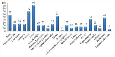
Background: Systemic lupus erythematosis (SLE) is a multisystemic chronic autoimmune disorder predominantly affecting women of child bearing age. The diverse clinical course and a high prevalence of SLE make it a major cause of morbidity and mortality especially in the younger population. Objective: To study the clinic Pathological profile of patients suffering from Systemic Lupus Erythematosus. Methodology: An Ambispective study was conducted at Kasturba Medical College and hospital, Manipal from January 2011 to December 2013. A total of One hundred twenty seven cases of SLE were classified as having SLE according to the revised American College of Rheumatology (ACR) classification criteria (1997). Results: The mean age of the study subjects in the present study was 29.85±12.61years.The male to female ratio was 1:11.7. Fever was the most common presenting symptom of the patients in the present study accounting to 92 out of total 127 cases studied (72.4%), followed by ascitis and skin rashes. Conclusion: SLE is a multisystem disorder affecting predominantly young females. Polyarthritis was the most common clinical feature. Incidence of  fever was the most common symptoms followed by dermatological manifestation .Hence all the cases of SLE should be evaluated in detail for the involvement of the all the systems and its manifestations in detail to improve the overall condition of the patients.

Keywords: SLE, Clinical, Ambispective, Rheumatology

Corresponding Author How to Cite this Article To Browse
Shwetha Patil, Post MD Tutor, Department of Pathology, Gadag Institute of Medical Sciences, Gadag, Karnataka, India.
Email:
Akshatha N, Patil S. A study to assess the clinic pathological profile of patients with systemic Lupus Erythematosus. Trop J Pathol Microbiol. 2019;5(7):493-499.
Available From
https://pathology.medresearch.in/index.php/jopm/article/view/294

Manuscript Received Review Round 1 Review Round 2 Review Round 3 Accepted
2019-07-10 2019-07-20 2019-07-25 2019-07-29
Conflict of Interest Funding Ethical Approval Plagiarism X-checker Note
No Nil Yes 9%

© 2019 by Akshatha N, Shwetha Patil and Published by Siddharth Health Research and Social Welfare Society. This is an Open Access article licensed under a Creative Commons Attribution 4.0 International License https://creativecommons.org/licenses/by/4.0/ unported [CC BY 4.0].

Introduction

There are several explanations for the origin of term lupus erythematosus. “Lupus” is Latin for wolf, and "erythro" is derived from Greek for "red". All explanations originate with the reddish, butterfly-shaped malar rash that the disease classically exhibits across the nose and cheeks[1]. The term lupus has also been mentioned by Rogerius Frugardi (1230 AD) to describe the erosive facial lesions and by Giovanni Manardi (1530 AD) to denote boils and ulceration of the lower extremity [2].

Systemic lupus erythematosis (SLE) is a multisystemic chronic autoimmune disorder predominantly affecting women of child bearing age. The diverse clinical course and a high prevalence of SLE make it a major cause of morbidity and mortality especially in the younger population. The prevalence of SLE has a vast demographic variation, with a higher incidece in the Africans and Hispanics. Although the incidence of SLE has drastically increased in the past decade due to the advancement in the diagnostic techniques, the prognosis has also improved significantly with an increase in survival rate especially due to early detection [3]. The pioneer in the study of lupus erythematosus in the modern era was Edmund L. Dubois. Then came the first lupus textbook, “Dubois' Lupus Erythematosus” by Dr. Dubois, a graduate of Johns Hopkins University.

Following this, the American College of Rheumatology Research and Education Foundation took up the task of recognizing an outstanding investigator in the field of systemic lupus erythematosus every year awarding them the Edmund L. Dubois Memorial Lectureship award. It was Dr. Dubois who recognised that SLE had no classic pattern and the diagnosis had to be based on an overall view of the entire clinical picture [3]. Being a prototypic autoimmune disease, SLE has a broad spectrum of clinical manifestations in association with autoantibodies to components of the cell nucleus. Its basic pathological features are that of inflammation and blood vessel abnormalities including band or occlusive vasculopathy, vasculitis, and immune complex deposition. “Autoantibody production” is the central immunologic disturbance in SLE. These antibodies interact with a host of self-molecules found in the nucleus, cytoplasm, or surface of cells. In addition the antibodies to soluble molecules such as IgG and coagulation factors are also present in the serum of the patients with SLE.

Because of the wide range of its antigenic targets, SLE is classified as a disease of “generalized autoimmunity” [5].

The characteristic pathology of SLE can be best explained with taking the example of the kidney which displays an array of features like increases in mesangial cells and mesangial matrix, inflammation, cellular proliferation, basement membrane abnormalities, and immune-complex deposits composed of IgM, IgG and IgA, as well as complement components. Other organ systems are also affected by SLE, but usually display non-specific inflammation or vascular abnormalities, although pathological findings are sometimes minimal [4].

SLE at its onset may involve one or more organ systems and additional manifestations may appear after a variable period of time. The systems involved in SLE are musculoskeletal, cutaneous, renal, nervous, hematological, vascular, pulmonary, gastrointestinal, and ocular. This is an observational study where we explore the various clinical presentations and pathological manifestations of SLE.

Objective

To study the clinic pathological profile of patients suffering from Systemic Lupus Erythematosus

Methodology

Study setting: Kasturba Medical College and hospital, Manipal.

Study duration: January 2011 to December 2013

Type of study: Ambispective Study

Sample size: A total of One hundred twenty seven cases of SLE were classified as having SLE according to the revised American College of Rheumatology (ACR) classification criteria (1997)

Sampling technique: Convenient sampling

Inclusion criteria

  1. All cases diagnosed and proved to be SLE for the first time at our institution in various departments of the hospital during the study period.
  2. Patients of all ages were included in the study.
  3. Patients were included in the study irrespective of the co morbidities present.

  1. Both inpatients and outpatients were included in the study.

Exclusion criteria

  1. All cases that were suspicious of SLE but did not satisfy the revised American College of Rheumatology (ACR) classification criteria [5].

Data collection and analysis: The details of the history of presenting complaints, associated symptoms, past medical history, findings on physical examination, reports of the various investigations done and procedures performed along with the follow up information were obtained from the files of the patients in the medical records department. Hematological and biochemical data was retrieved by using hospital and laboratory information system.

All patients with a diagnosis of SLE were evaluated, along with the details of history, physical examination and clinical examination was performed. All the information were entered in excel sheet and then analyzed using SPSS v 20.

Results

A total number of 127 cases of SLE were included in the present study and their clinical profile were analyzed.

Table-1: Social profile of the study subjects.

Social profile Frequency Percentage
Age < 21 Years 32 25
21-40 Years 68 54
>40 years 27 21
Gender Male 10 8
Female 117 92

The age of the patients in the present study ranged from 6 to 63 years. The mean age of the study subjects in the present study was 29.85±12.61years. Maximum number of patients (68/127 i.e. 54%) fell in the age group of 21-40 years. The male to female ratio was 1:11.7, showing a female predominance (117/127 i.e. 92%).

patho_294_01.jpg

Figure-1: Clinical Profile of the study subjects

Fever was the most common presenting symptom of the patients in the present study accounting to 92 out of total 127 cases studied (72.4%), followed by ascitis and skin rashes.

Musculoskeletal manifestations: Seventy five patients out of 127 cases studied (59%) had musculoskeletal manifestations at presentation. Arthritis was a common presenting symptom accounting to 70/127 cases (55.1%). Small joints were involved predominantly and were associated with pain, occasionally accompanied by morning stiffness and swelling. Other musculoskeletal manifestations included myalgia and muscle weakness.

Mucocutaneous manifestations: Seventy nine out of 127 cases presented with mucocutaneous manifestations accounting to 62.2%. The dermatological manifestations included rashes (papulosquamous and annular lesions) which were present in 58 out of 127 cases, (45.7%) usually involving the distal extremities. Malar rash was seen in 34/127 patients (26.8%) and were usually accompanied by pruritis. Raynaud phenomenon was noted in four cases while two patients had gangrene of the toes. Other manifestations included alopecia which was seen in 25/127 cases (19.7%). Twenty four cases showed photosensitivity accounting to 18.9%. Oral ulcers were seen in 26/127 (20.5%) of patients, while bruising and ecchymosis were seen in 22/127 (17.3) patients.

Skin immunofluorescence: Immunofluoresence of the skin lesions was done in 34/127 cases (26%), of which 31/34 (91.1%) cases tested positive for lupus band, hence suggesting SLE while 3 cases (9%) did not show lupus (Chart 4).

Skin biopsies were sent for hispathological analysis in 29 cases and all the 29 had features suggestive subacute and chronic cutaneus lupus erythamatosus. The biopsies showed changes like hyperkeratosis, basal cell layer vacuolation, subepidermal, peri-vascular and peri-appendiceal inflammatory infiltrate, epidermal atrophy etc. ANA profile was done in 24 of the 29 cases, 15 of which showed positivity to anti RNP-Sm antibodies.

Cardiovascular manifestations: Twenty four out of the 127 cases (18.8%) had cardiovascular manifestations at presentation, which included valvular incompetence in 17 cases (70%) mostly involving the mitral and aortic valve, pericardial


effusion in 3 cases (12%) without any sign of tamponade and myocarditis in 2 cases (8%) as seen on ECHO. No significant ECG changes were observed.

Pleuropulmonary manifestations: Nineteen of the 127 cases (14.9%) had pleuropulmonary involvement at presentation. Sixteen (12.5%) had pleural effusion. Four cases had pneumonia, while two had pleuritic rub. Other manifestations included breathlessness, cough, bronchiectasis, etc.

Renal manifestations: A total of 58 cases (45%) showed renal involvement at presentation. The patients presented with symptoms such as reduced urinary output and facial puffiness. On investigation, 48 cases (82.7%) had significant protienuria and 14 cases (24%) showed deranged renal function tests (elevated serum urea and creatinine). Four cases showed urinary sediments/ casts and 12 cases (20%) had grade 1 – 2 renal parenchymal changes on ultrasound.

Lupus nephritis: Renal biopsies were done in 38 cases (65.5%) of which 36 cases (94.73%) proved to be lupus nephritis. These cases were then sub-classified into different classes of lupus nephritis and their reports are represented in chart 5. The remaining 2 biopsies were inadequate for opinion.

It was seen that majority of the cases showed the histological pattern of diffuse proliferative glomerulonephritis hence belonged to lupus nephritis – class 4 (18/38 i.e. 47.38%), followed by focal segmental glomerulo nephritis – class 3 (7/38 i.e., 18.42%), mesangio proliferative glomerulo nephritis – class 2 (5/38 i.e. 13.15%), membranous glomerulonephritis – class 5 (4/38 = 10.52%) and minimal change disease – class 1 (2/38 = 5.5%).

Organomegaly & ascitis: Hepatomegaly was a common finding, seen in 42/127(33.1%) cases, followed by splenomegaly which was seen in 21/127 (16.5%). Ascitis was observed in 16 cases (12.6%).

Neuropsychiatric manifestations: Twenty two patients (17.33%) presented with neurological symptoms, which included altered sensorium in 6 cases, loss of power in 5 cases, seizures (new onset) in 3 cases, depression in 2 cases and other manifestations like hemiparesis, neuropathy, involuntary movements and dysarthria (1 case each). Imaging was done in 8 cases which showed hydrocephalus in 2 cases, meningeal enhancement in 2 cases, cerebral atrophy in one case and acute lacunar infarcts in one case.

Two cases showed a normal imaging study. Nerve conduction study was done in 2 cases which was normal.

Ocular manifestations:Two patients showed ocular manifestations presenting as photophobia and loss of vision. On examination one patient showed papilledema and the other showed retinal hemorrhage, who, when further evaluated also showed central retinal artery occlusion.

Menstrual Irregularity and Recurrent

Abortions:Among the 117 female patients studied, 5 patients aged 6yrs, 10yrs, 11 yrs (two patients) and 12yrs, had not attained menarche. Menstrual irregularities were seen in 10/112 (8.9%) patients, which included menorrhagia in 9 patients and polymenorrhea in one patient.

Seven patients gave history of recurrent abortions of which 4 were positive for antiphospholipid antibodies. These patients were observed to have IgG anticardiolipin antibody.

A total of 36 cases were diagnosed as lupus nephritis by biopsy. All these patients had significant protienuria and other renal symptoms like oliguria, and renal parenchymal changes on ultrasonography.

Ten cases were diagnosed with CNS lupus, of which 3 cases had new onset seizures, where other related causes were ruled out. Imaging studies were done in all the cases which showed features like meningeal enhancement, cerebral atrophy and hydrocephalus.

Six patients were diagnosed with Antiphospholipid antibody syndrome. All 6 cases were female patients with menstrual irregularities and 3 cases with history of recurrent abortions. Three of these cases showed DRVVT positivity, while the other three showed IgG positivity to Anticardiolipin antibody.

Seven cases were diagnosed with ITP. All these cases presented with thrombocytopenia with anemia and no associated clinical features suggestive of other causes. Anti platelet antibody was done in 4 of these cases, of which 2 were positive. Bone marrow aspiration and biopsy was done in 2 cases of which one case showed erythroid dyspoiesis and one case showed increase in the megakaryocytes.

Fourteen of the 127 patients had diabetes mellitus, with their age ranging from 35 – 57 yrs, of whom one patient aged 57 yrs and was an old case of diabetes while 13 were newly diagnosed.


Of these 13 cases two were diagnosed with type 2 diabetes mellitus and rest 11 cases were steroid induced diabetes. Eleven of the 127 patients were hypertensive, of which 7 cases were old cases and rest 4 were newly diagnosed, all of whom were females. Of the 4 cases that were newly diagnosed, 3 had lupus nephritis.

Discussion

This was a retrospective-prospective study of the clinical and hematological profile of patients with systemic lupus erythematosus at a tertiary medical centre for a period of 3 years. The clinical manifes-tations of all the cases in comparison with other studies are depicted in the below.

SLE being a disease predominantly effecting the female population, a female preponderance in the patients was an expected result in the study. The male of female ratio was observed to be 1:11.7, and was in concordance with other studies by Mok et al[6], Sasidharan et al [7], Voulgareli et al [8], Fathi et al [9] got a relatively higher M:F of 1:44.

Similarly, the median age of presentation at 29.5 years in the present study, is also expected as SLE is a disease affecting the women of childbearing age group and hence is in concordance with the other studies[10], [11]. However in the study by Voulgareli et al [8], the mean age was 42yrs, which is much higher than the present study.

Unlike the other studies conducted by authors like Mok et al [6](82%), Binoy et al [12] (89.3%), Aleem et al [11] (80.4%) and Paudyal et al[13] (93%) where arthritis was the most common presenting symptom among the patients, followed by fever; the present study reported fever as the most common presenting symptom among the patients occurring in 72.4% of the cases, followed by skin manifestations (62.2%) and arthritis which was seen in 55.1% of the cases.

This was in concordance with a study by Agarwal et al [10] who also reported fever as the most common presenting symptom accounting to 82.8%. The incidence of arthritis in the present study was in concordance with the studies by Voulgareli et al[8] (50%), Fathi et al [9] (60%), Agarwal et al [10] (52.9%) and Kosaraj et al [14](64.58%).

Dermatological manifestations occurred in 62.2% of the cases in the present study and closely matched with the study conducted by Aleem et al [11]

(64.3%), as against 91.7% in a study by Saigal et [15], and 97% in the study by Paudyal et al [13], while it was seen in only 18.5% in another study by Sasidharan et al [7]. Among the lupus specific cutaneous lesions, the malar rash was seen in 26.8% of the patients as opposed to all the other studies by Agarwal et al[10], Aleem et al[11], Paudyal et al[13] and Kosaraj et al[14] where the incidence of malar rash was at seen in a minimum of 35% Kosaraj et al[14] of the patients upto76.76 % Fathi et al [9].

Unlike the study by Sultan S M et al [16], where there is a clear increase in the incidence in discoid lupus in male patients, in the present study all cases (4.7%) who had discoid lupus were female patients. The incidence was much higher in the studies by Agarwal et al [10] (32.2%), Paudyal et al [13] (23%) and Fathi et al [9] (22.7%).

Photosensitivity was less frequently seen in the cases in the present study accounting for 24cases (18.9%), whereas it has been noted in a relatively higher frequency accounting to 40%, 44.86%, 32%, 75%, 34%, 45%, 63.2%, 29% and 27.08% of the cases in studies by Voulgareli et al [8], Fathi et al [9], Binoy et al [12] and Kosaraj et al [14] respectively.

Oral ulcers were seen in 20.5% of the cases in the present study at presentation, which is in concordance with the studies by Voulgareli et al [8] and Kosaraj et al [14] which showed an incidence of 20% and 25% respectively as opposed to the studies by Mok et al[6] and Swaak et al [17] where it was encountered less frequently in only 6% and 9% cases respectively. A relatively higher frequency, i.e., 64% and 61.7% cases of oral ulcers were seen at presentation in the studies by Binoy et al [12] and Saigal et al [15] respectively.

Alopecia was seen in 19.7% of the cases in the present study, which is in concordance with the studies conducted by Kosaraj et al[14](18.5%) and Swaak et al[17](16%), while higher incidence of alopecia were seen in other studies by Binoy et al [12] (60%) and Saigal et al [15] (65%).

Raynaud phenomenon was less frequently encountered in our cases accounting for 3.1% of the cases, which is in concordance to a study conducted by Binoy et al [12] and Fathi et al [9], who reported Raynaud phenomenon in 2.7% and 2.16% of the cases respectively. While a higher incidence of Raynaud phenomenon was documented in other studies by Saigal et al [15] (21.7%), Mok et al [6]


(10.5%) and Swaak et al [17] (46%).

Among the organ involvement, the kidney was the most frequent organ involved inthe patients of the present study, with 45% of the patients presenting with renal manifestations, which is in concordance with other studies by Mok et al [6], Sasidharan et al[7], Voulgareli et al [8], Swaak et al[17], Agarwal et al [10], Aleem et al [11], and Fathi et al [9] who also reported kidney as the most frequent organ involved accounting to about 27%, 23%, 12.5%, 87.03%,33.3%, 56.7%, 26%, 20.83% respectively.

The renal manifestations included protienuria, deranged RFT and urinary casts and sediments which were seen in 37.7%, 88.88% and 9.4% of the cases respectively. This is in concordance with a study by Agarwal et al[10] who reported 7.15% cases of protienuria, 24.1% cases of deranged RFT and urinary casts and sediments in 3.4% cases.

Among the 38 study patients who underwent renal biopsy, 36 were confirmed to be lupus nephritis. Of the 36 cases of lupus nephritis, most of the cases were classified as Class IV (18/38 = 47.38%), and Class III LN (7/38 = 18.42%), followed by Class II (5/38 = 13.15%), and Class V (4/38 = 10.52%). Only 2 cases (5.5%) showed minimal change disease – class 1.

This is to a certain extent is similar to the series by Mok et al[6] and Saigal et al[15], who found the overall frequency of Class II, III, IV and V is 1%, 20%, 65%, 14% and 0%, 5%, 90%, 5% respectively.

Pleuropulmonary manifestations were seen in 14.9% of the cases, which included pleural effusion, pleuritis and pneumonitis, which is in concordance with the study by Saigal et al [15], Fathi et al [9], Aleem et al [11] and Kosaraj et al[14] who reported 11.7%, 15.16% 15.9% and 12.5% of the cases respectively, while other studies by Sasidharan et al [7] and Binoy et al [12] showed a lower frequency of pulmonary manifestations (3% and 8% respectively), and a higher incidence of 20% was seen in the study by Paudyal et al [13].

Among the pleuropulmonary manifestations documented in the present study, pleural effusion was the most common manifestation which in concordance with the other studies as well.

Cardiovascular manifestations were seen in 18.8% of the cases in the present study, which included valvular deformities, pericarditis, myocarditis, and pericardial effusion.

Similar to the present study the studies by Aleem et al [11], Fathi et al [9], Paudyal et al [13] and Swaak at al [17] also documented 20.8%, 24.86%, 13% and 15% cases with cardiovascular involvement respectively. A lower incidence of 5.3% and 6.7% was seen in studies by Binoy et al [12] and Saigal et al [15] respectively.

The incidence of neuropsychiatric manifestations in the cases of the present study was relatively high, accounting to about 17.33%, which is in concordance with other studies by Voulgareli et al [8], Binoy et al [12] and Saigal et al [15] which showed an incidence of 17.5%, 13.3% and 13.3% respectively. A lower frequency was observed in the studies by Mok et al [6], Sasidharan et al [7], Agarwal et al [10] and Swaak et al [17] which was 4%, 4%, 3.5% and 6% respectively.

Other manifestations included hypothyroidism which was seen in 12 patients (9.54%) in the present study which is in concordance with the studies done by Sasidharan et al[7], Saigal et al[15] and Paudyal et al[13] which was 12%, 8% and 11% respectively. Ocular manifestations which included visual distur-bances caused by retinitis and papilledema was seen in 2 patients in the present study accounting to 1.5%, which is in concordance with the studies done by Sasidharan et al[7] and Paudyal et al [13] who reported 3 cases (3%) and 1 case (1%) respectively.

Serositis which included pleural effusion and pericardial effusion were seen in 22 patients accounting to 17% of the cases in the present study, which is in concordance with Mok et al [6] who reported 26 cases (16%), while Agarwal et al [10] reported a relatively lesser number of cases (8 cases – 9.2%) and Voulgarelis et al [8] reported a higher number of cases (11 cases – 28.5%).

Cutaneous vasculitis was seen in a total number of 9 patients in the present study accounting to 7%, which was low in comparison with 32 cases (18%) and 70 cases (37.84%) in the studies by Mok et al[6] and Fathi et al[9] respectively. Pudyal et al[13] reported a similar number of 5 cases accounting to 7% in their study.

Limitation of the study: Investigations such as Skin Biopsy Immunofluorescense and Renal Biopsy were not performed in few patients as they were not willing to get it done. Few patients were lost to follow up, hence the pathological changes could not be assessed


Conclusion

The clinical features of the SLE patients seen in the present study cannot be generalized to the whole community and hence it cannot be taken as a true representative of the clinical profile of SLE Patients. The follow-up of these SLE patients was not available; hence, the change in clinical profile of these patients as the disease progress was not observed.

SLE is a multisystem disorder affecting predominantly young females. Polyarthritis was the most common clinical feature. Incidence of fever was the most common symptoms followed by dermatological manifestation. Kidney was the major system involved among majority of the cases followed by cardiac, Neuropyschiatric and Pulmonary. Hence all the cases of SLE should be evaluated in detail for the involvement of the all the systems and its manifestations in detail to improve the overall condition of the patients.

What the present study adds to the existing knowledge?

SLE being a multi-systemic disorder presents with a large spectrum of symptoms, a complete evaluation of the patient including detailed hematological as well as biochemical and immunological studies should be done for every patient to detect early lesions and hence improving the prognosis.

Author’s contribution

The data collection and analysis was performed by Dr. Akshatha N, The review of articles, introduction, references and Manuscript preparation was done by both Dr. Akshatha N and Dr. Shwetha Patil. The final proof reading was done by Dr. Shwetha Patil.

Reference

01. Mallavarapu RK, Grimsley EW. The history of lupus erythematosus. South Med J. 2007;100(9)896-898. doi:10.1097/SMJ.0b013e318073c9eb [Crossref][PubMed][Google Scholar]

02. Blotzer JW. Systemic lupus erythematosus I- historical aspects. Md State Med J. 1983;32(6)439-441. [Crossref][PubMed][Google Scholar]

03. Rus V, Maury EE, Hochberg MC. The epidemiology of systemic lupus erythematosus, In- Dubois' Lupus Erythematosus, Wallace DJ, Hahn BH (Eds). Lippincott Williams and Wilkins, Philadelphia. 2002. [Crossref][PubMed][Google Scholar]

04. George Bertsias, Ricard Cervera D. Systemic Lupus Erythematosus- Pathogenesis and Clinical Features. 5th ed, London NW1 7BY, UK Elsevier. 2011;476–505. [Crossref][PubMed][Google Scholar]

05. Hochberg MC. Updating the American College of Rheumatology revised criteria for the classification of systemic lupus erythematosus. Arthritis Rheum. 1997;40(9)1725. doi: 10.1002/art.1780400928 [Crossref][PubMed][Google Scholar]

06. Mok CC, Lee KW, Ho CT, Lau CS, Wong RW. A prospective study of survival and prognostic indicators of systemic lupus erythematosus in a southern Chinese population. Rheumatology (Oxford). 2000;39(4)399-406. doi: 10.1093/rheumatology/39.4.399 [Crossref][PubMed][Google Scholar]

07. Sasidharan PK, Bindya M, Sajeeth Kumar KG. Hematological manifestations of SLE at initial presentation- is it underestimated?. ISRN Hematol. 2012. doi: 10.5402/2012/961872 [Crossref][PubMed][Google Scholar]

08. Voulgarelis M, Giannouli S, Tasidou A, Anagnostou D, Ziakas PD, Tzioufas AG. Bone marrow histological findings in systemic lupus erythematosus with hemato-logic abnormalities- a clinicopathological study. Am J Hematol. 2006;81(8)590-597. doi: 10.1002/ajh.20593 [Crossref][PubMed][Google Scholar]

09. Fathi H, Ahmed EF, Abdelgawad MM, Elbayoumi Y. Cutaneous manifestations of systemic lupus erythematosus - a retrospective study from Egypt. Gulf J dermatology Venerol. 2010;17(1)36–42. [Crossref][PubMed][Google Scholar]

10. Agrawal S, Jain A, Rajput A, Tiewsoh I. A cross-sectional hospital based study of clinical and immunological profile of systemic lupus erythematosus patients from central rural India. Indian J Allergy, Asthma Immunol. 2013;27(1)33. doi: 10.4103/0972-6691.116614 [Crossref][PubMed][Google Scholar]

11. Aleem A, Al Arfaj AS, khalil N, Alarfaj H. Haematological abnormalities in systemic lupus erythematosus. Acta Reumatol Port. 2014;39 (3)236-41. [Crossref][PubMed][Google Scholar]


12. Binoy JP, Muhammed F, Kumar N, Razia MV. Clinical profile of systemic lupus erythematosus in North Kerala. J Indian Rheumatol Assoc. 2003;11;94-7. [Crossref][PubMed][Google Scholar]

13. Paudyal BP, Gyawalee M. Clinical profile of patients with systemic lupus erythematosus. JNMA J Nepal Med Assoc. 2012;52(187)111-7. [Crossref][PubMed][Google Scholar]

14. Kosaraju K, Shenoy S, Suchithra U. A cross-sectional hospital-based study of autoantibody profile and clinical manifestations of systemic lupus erythema-tosus in south Indian patients. Indian J Med Microbiol. 2010;28(3)245-7. doi: 10.4103/0255-0857.66487 [Crossref][PubMed][Google Scholar]

15. Saigal R, Kansal A, Mittal M, Singh Y, Maharia HR, Juneja M. Clinical profile of systemic lupus erythematosus patients at a tertiary care centre in Western India. J Indian Acad Clin Med. 2011;13;27-32. [Crossref][PubMed][Google Scholar]

16. Sultan SM, Begum S, Isenberg DA. Prevalence, patterns of disease and outcome in patients with systemic lupus erythematosus who develop severe haematological problems. Rheumatology (Oxford). 2003;42(2)230-4. doi: 10.1093/rheumatology/ keg069 [Crossref][PubMed][Google Scholar]

17. Swaak AJ, Van den Brink, HG, Smeenk, RJ, Manger K, Kalden JR, Tosi S, Smolen JS. Systemic lupus erythematosus: clinical features in patients with a disease duration of over 10 years, first evaluation. Rheumatology(Oxford, England). 1999;38(10)953–958. [Crossref][PubMed][Google Scholar]