E-ISSN:2456-1487
P-ISSN:2456-9887
RNI:MPENG/2017/70771

Research Article

Diagnosing

Tropical Journal of Pathology and Microbiology

2019 Volume 5 Number 7 July
Publisherwww.medresearch.in

Utility of nested PCR in diagnosing extra-pulmonary Tuberculosis

Kaur Gill M.1*, Khanna A.2, Sharma S.3
DOI: https://doi.org/10.17511/jopm.2019.i07.05

1* Manmeet Kaur Gill, Associate Professor, Department of Microbiology, Sri Guru Ram Das Institute of Medical Sciences and Research, Amritsar, Punjab, India.

2 Ashish Khanna, Professor, Department of Microbiology, Sri Guru Ram Das Institute of Medical Sciences and Research, Amritsar, Punjab, India.

3 Sarbjeet Sharma, Professor and Head, Department of Microbiology, Sri Guru Ram Das Institute of Medical Sciences and Research, Amritsar, Punjab, India.

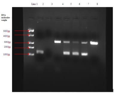
Background: Tuberculosis has always been a burden for developing countries such as India. Conventional techniques (smear microscopy and culture) used for diagnosing this highly contagious disease have a lower sensitivity in comparison to the advanced diagnostic techniques. Polymerase chain reaction has emerged as a better alternative with higher specificity and sensitivity. This study was therefore designed for evaluating the efficacy of IS6110 sequence based nested polymerase chain reaction (nPCR) in detecting Mycobacterium tuberculosis (MTB) in extra-pulmonary clinical samples. Material and Methods: The study comprised of 195 samples of extra-pulmonary origin obtained from the patients with history suggestive of tuberculosis. The samples were immediately processed and subjected to Ziehl-Neelsan (ZN) staining for presence of acid fast bacilli (AFB), inoculation on Lowenstein-Jensen (LJ) medium for culture and nested PCR using specific primers that target the 123bp fragment of IS6110 of the MTB- complex. Results: It was observed that 72/195 (36.92%) samples were diagnosed positive for TB using PCR whereas only 29/195 (14.87%) were positive by smear and 32/195 (16.41%) by culture on LJ. Total positive samples by conventional methods (either smear or culture) were 35/195 (17.94%). These results clearly indicated that PCR had higher sensitivity over conventional methods with a significant difference in p value (p<0.05). Conclusions : It was concluded from the study that nPCR method targeting IS6110 gene sequence is more efficient, rapid, sensitive, and specific method for detecting Mycobacterium tuberculosis genetic material in extra-pulmonary tuberculosis specimens of patients with suspected history of TB in contrast to the available conventional methods.

Keywords: Mycobacterium tuberculosis, Conventional methods, Nested polymerase chain reaction, IS6110 sequence

Corresponding Author How to Cite this Article To Browse
Manmeet Kaur Gill, Associate Professor, Department of Microbiology, Sri Guru Ram Das Institute of Medical Sciences and Research, Amritsar, Punjab, India.
Email:
Kaur Gill M, Khanna A, Sharma S. Utility of nested PCR in diagnosing extra-pulmonary Tuberculosis. Trop J Pathol Microbiol. 2019;5(7):443-448.
Available From
https://pathology.medresearch.in/index.php/jopm/article/view/286

Manuscript Received Review Round 1 Review Round 2 Review Round 3 Accepted
2019-06-26 2019-06-04 2019-07-10 2019-07-16
Conflict of Interest Funding Ethical Approval Plagiarism X-checker Note
No Nil Yes 9%

© 2019 by Manmeet Kaur Gill, Ashish Khanna, Sarbjeet Sharma and Published by Siddharth Health Research and Social Welfare Society. This is an Open Access article licensed under a Creative Commons Attribution 4.0 International License https://creativecommons.org/licenses/by/4.0/ unported [CC BY 4.0].

Introduction

Airborne infections are a serious threat to the people of developing countries. Larger population, poor standards of living and hygiene, and major use of public transport in these countries make this infection more feasible. Tuberculosis (TB) caused by Mycobacterium tuberculosis is a major air borne infectious diseases. The most common clinical presentation of this disease is pulmonary tuberculosis, but surprisingly the extra-pulmonary tuberculosis (EPTB) has emerged as an equally grave clinical problem.

Interestingly, the number of patients presenting with EPTB has increased to 15 to 20 per cent among all the tuberculosis patients and the number is even higher among HIV positive patients (50%) [1]. The diagnosis of EPTB using the conventional methods has always been a problem due to lower sensitivity of the microscopy and culture, risk of contamination due to long incubation period and higher false negative results.

Literature survey has revealed acid fast bacilli smear positivity is 10 to 37% TB patients, while the culture positivity varies from 12 to 80% [1]. There is lack of standardization of the methodology among various diagnostic laboratories as they generally utilize same methodologies without researching on new alternatives.

In contrast to these conventional methods of diagnosing TB, new and improved methods that focus on nucleic acid-based diagnosis have been developed. These newer diagnostic methods are more reliable, with shorter turnaround time, higher sensitivity and specificity for detection and identification of Mycobacterium tuberculosis in pulmonary as well as extra pulmonary samples of TB patients [2, 3]. Among these, the Polymerase Chain Reaction (PCR) has emerged as a better diagnostic tool for EPTB.

It has a higher sensitivity and specificity and has been evaluated using a number of primer targets such as TRC4, devR, 65 Kda, IS6110, etc [4-8]. The present study was therefore designed to compare the conventional diagnostic methods with nested Polymerase Chain Reaction (nPCR) using primer against IS6110, for diagnosing EPTB in the patients visiting a tertiary care hospital of North India. after obtaining ethical approval from the ethical committee of the institution.

Material and Methods

Ethical approval: The study was carried out after obtaining ethical approval from the ethical committee of the institution.

Study design & data collection: It was a prospective study carried out over a period of five years.

Settings: The study was designed and carried out in the Department of Microbiology, S.G.R.D Institute of Medical Science and Research, a tertiary care hospital of North India.

Sample size: The study comprised of non-repeat extra-pulmonary samples from 195 patients, presenting with clinical signs and symptoms suggestive of tuberculosis. The samples from these patients comprised of 60 samples of ascitic fluid, 52 pleural fluid, 23 cerebrospinal fluid, 14 lymphnode aspirates, 13 endometrial biopsies, 12 pus samples, 11 synovial fluid samples, and 10 urine samples. A brief clinical history of the illness was also recorded.

Inclusion criteria- The patients of both sexes and all age groups attending the outpatient department of the hospital with suspicion of tuberculosis from clinical history and radiological evidence were included in the study. It was ensured that none of the patient had received any anti-tubercular treatment for more than four weeks.

Exclusion criteria

The following patients were not included in the study:

  • Those who had been diagnosed and treated for tuberculosis.
  • Those who had been put on anti-tubercular drugs but the treatment failed.
  • Those who were defaulters during the treatment schedule for tuberculosis.
  • Those patients who had relapse of tuberculosis after the completion of treatment.

Sample processing

Conventional methods- The patient samples received in the department of Microbiology were apportioned for conventional methods and PCR procedure. Each clinical sample was subjected to smear microscopy, one direct and the other after the required concentration. The concentration of the samples was made as per the standard methods and the nature of samples [9].


Fluid samples were centrifuged for 30 minutes at 3000 rpm before analysis, tissue biopsy samples were homogenized, and the pus samples were processed using the N-acetyl-L-cysteine NaOH method. The lymph node aspiration samples obtained aseptically were processed directly. The processed samples were then stained using Ziehl-Neelsen (ZN) staining to look for acid fast bacillus.

The samples were inoculated on Lowenstein-Jensen (LJ) medium following standardbacteriological procedures for the culture of Mycobacterium tuberculosis [10]. Briefly, the processed sample was directly inoculated on LJ medium in duplicate.

The seeded media was bacterial growth was examined daily for bacterial growth during the first week for detecting rapidly growing M. tuberculosis or for the presence of any contamination. After the initial week, the bacterial cultures were examined on weekly basis for 8 weeks. After the completion of 8 weeks, the bacterial cultures were discarded. Mycobacterium tuberculosis growth on the cultures was identified on the basis of a relatively slow growth rate, lack of pigmentation in the colonies and a positive Niacin test.

Polymerase chain reaction: PCR for MTB were performed using IS6110 primers specific for the same.

DNA extraction from clinical samples: A portion of material obtained after concentration was then further processed for DNA extraction using commercially available kit viz. Amplification Reagent Set for Mycobacterium tuberculosis, Bangalore Genei, Bangalore, India following manufacturer’s guidelines

Nested polymerase chain reaction (nPCR): Polymerase chain reaction thermal cycler and single tube nPCR along with the primer targeting IS6110 gene sequence of M. tuberculosis was used for diagnosing EPTB from processed clinical samples.

The primers were purchased from Bangalore Genei, Bangalore, India. Two sets of primers were used; first primer used was for 220 bp sequence of IS region of Mycobacterium tuberculosis-complex DNA sequence. This sequence was amplified during the first round of amplification. In the second step, the nested primer was added to the mixture for amplifying the 123 bp amplification product.

Gel electrophoresis of the DNA obtained after amplification: The amplified DNA product was separated using gel electrophoresis.

The gel used in this experiment was agarose gel having a concentration of 2.5% and mixed with ethidium bromide. The electrophoresis was performed at 110 volts and the gel was then visualized using the UV transilluminator. All the PCR reactions were carried out along with positive and negative controls for avoiding any risk of cross-contamination and preventing any false positive reactions.

Statistical analysis: SPSS version 17.0 software and MS excel 2007 were used for statistic analysis. Culture on LJ medium was used as gold standard and sensitivity and specificity of PCR was calculated based on that.

Proportion test was used for determining the level of significance among the compared methods. The significant value chosen for the study was a probability value less than 0.05 (p< 0.05).

Results

Microbiological study- The study was carried out on 195 non repeat extra-pulmonary samples which were processed for diagnosing Tuberculosis.The study included conventional methods of analysis by smear microscopy using Ziehl-Neelsen staining and culture on Lowenstein-Jensen medium as well as Molecular method using nested polymerase chain reaction approach.

It was observed that 72/195 (36.92%) samples were diagnosed positive for TB using PCR whereas only 29/195 (14.87%) were positive by smear microscopy and 32/195 (16.41%) by culture on LJ. Total positive samples by conventional methods (either smear or culture) were 35/195 (17.94%) (Table 1).

patho_286_01.jpg


Line 1 DNA molecular weight marker

Line 2 is Positive control (123bp)

Line 3 is Negative control (no band)

Line 4 Sample 1 is showing no band at 123 bp therefore result is negative

Line 5 Sample 2 is showing band at 123bp, so the result is positive

Line 6 Sample 3 is showing band at 123bp, so the result is positive

Line 7 Sample 4 is showing band at 123bp, so the result is positive

Line 8 Sample 5 is showing band at 123bp, so the result is positive

Among the conventional methods, only 9 smears were found to be positive for acid fast bacilli by ZN stain before concentrating the samples and a total of 29 samples were found to be positive after concentration procedure (Table 1).


Table-1: Results of conventional bacteriological tests and PCR.

Samples Smear pos/samples investigated Culture pos/ samples investigated Smear/culture pos/samples investigated PCR pos/ samples investigated
Ascitic fluid 9/60 10/60 11/60 25/60
Pleural fluid 3/52 4/52 6/52 14/52
CSF 0/23 0/23 0/23 0/23
Synovial fluid 0/11 0/11 0/11 6/11
Endometrial biopsy 0/13 0/13 0/13 3/13
Urine 4/10 4/10 4/10 6/10
Pus 6/12 6/12 6/12 8/12
Lymphnode aspirates 7/14 8/14 8/14 10/14
Total 29/195 32/195 35/195 72/195

All among 29 samples which were found to be positive by microscopy also showed growth on the LJ medium except 3 samples of ascitic fluid which did not show growth even after incubating for 8 weeks. However 6 additional samples which showed negative result on microscopic analysis, were positive for growth on the LJ medium. Total positive samples by conventional methods (either smear or culture) were 35/195 (17.94%) (Table 2).

Table-2: Comparison of results of direct microscopy, culture and PCR (N=195)

Direct Smear Microscopy Growth on LJ medium PCR
Positive Negative Positive Negative
Positive 29 26 3 29 0
Negative 166 6 160 43 123
Total 195 32 163 72 123

Polymerase chain reaction- Polymerase chain reaction was performed on all the 195 samples using primers targeting IS6110. Among the samples analyzed, 72 samples were found to be positive using the PCR method (Table 1). The samples which were detected positive using the conventional methods were also found positive using the PCR

technique (Table 2). This technique was able to detect 37 additional positive samples in contrast to the conventional method.

The PCR technique showed the highest positive results (36.92%), followed by the culture method (16.41%) and the least sensitive test was the direct microscopy method (14.87%).

Discussion

Tuberculosis is a preventable infectious disease caused by Mycobacterium tuberculosis. The effective management of this disease is dependent on accurate diagnosis of the infectious agent. The conventional method such as direct smear microscopy has an advantage of providing earlier diagnosis, but lacks sensitivity as it is dependent on the presence of sufficient number of bacteria in the sample and extra pulmonary samples usually are paucibacillary in nature. There are several problems experienced while analyzing EPTB samples. Some of these being lower microbial count in the sample, insufficient sample, and non-uniform distribution of microbes in the sample.


Consequently, conventional methods are not suitable for diagnosing tuberculosis from extra-pulmonary samples; necessitating requirement of advanced techniques such as PCR. The latter is a superior diagnostic method that provides accurate and rapid results with high sensitivity and specificity. The PCR can be performed using a large number of gene targets of Mycobacterium tuberculosis. There are many primers available for the amplification of gene targets such as 65 Kda, IS6110, devR, TRC4 etc. The PCR technique used in this study included the primer IS6110, specific for Mycobacterium tuberculosis complex [13]. The IS6110 insertion sequence was amplified for obtaining accurate results since most stains of Mycobacterium tuberculosis carry10-15 copies of this insertion sequence.

This study conducted on 195 samples showed interesting results. It was observed that the conventional methods showed positive results in much fewer numbers of samples in contrast to the PCR technique. The culture positivity was observed in32/195 (16.41%) samples, and the smear method showed positive results in 29/195 (14.87%) samples. In contrast, the PCR technique showed positive results in 72/195 (36.92%) samples.

The results of conventional methods were compared with those of the PCR technique and a statistical significant difference was observed among the results (p≤0.015) (Table1). Further PCR technique showed 100% sensitivity as compare to the gold standard i.e. culture on LJ. A study conducted by Tiwari et al showed that PCR technique was efficient in identification of 62% positive results among the EPTB samples, and among the results 57% were found to be AFB negative [11]. The studies by Gill MKet aland Kesarwani et al reported that PCR technique using IS6110 sequence was more effective in diagnosing tuberculosis in EPTB samples [12, 13].

A similar study by Negi et al compared the PCR techniques using different primers, and it was observed that IS6110 showed better results (77%) in contrast to other gene targets like 38 kDa (72%), 85B protein (73%) and 65kDa (75%). Several studies have shown that the use of IS6110 as gene target during PCR analysis provides reproducible results using simpler methodology. A study by Sekar et al (2008) has shown similar results of better sensitivity and reproducibility of IS6110 gene sequence [1].

However, the studies by Daset al (1995) and D.S Chauhan et al(2007) have reported absence or the presence of fewer copies of target sequence IS6110, in some strains of Mycobacterium tuberculosis related to geographical origin [14,15]. The isolates from Indian subcontinent usually have less copies of IS 6110 as compared with 8-15 found in strains from most of the developed countries. Thus presence of variable number of target gene sequence in some of the strains of Mycobacterium tuberculosis makes it necessary to evaluate PCR protocols using different gene sequences as per the region of study.

Limitations: In this study we used primers against gene sequence IS 6110 but since the number of copies of the target sequence is an important determinant of PCR sensitivity and isolates from Indian subcontinent usually contain less number of copies of IS6110 so it becomes necessary to evaluate PCR protocol based on other genes sequences of MTB also.

Conclusions

The present study provides strong evidence that use of nPCR targeting IS6110 gene sequence is a simple, rapid, accurate and highly sensitive method for diagnosing Tuberculosis in extra-pulmonary samples compared to the conventional methods. To conclude we can say that nPCR assay targeting IS6110 is highly useful in the establishment of diagnosis of EPTB where there is strong clinical suspicion. However, further studies are necessary to evaluate the samples having less target gene copies specific to the geographical region of study. In such cases use of multiplex PCR employing multiple target gene sequence should be utilized.

What does this study add to existing knowledge?

The present study provides strong evidence that the use of nPCR targeting IS6110 gene sequence is a simple, rapid, accurate and highly sensitive method for diagnosing Tuberculosis in extra-pulmonary samples compared to the conventional methods.

Author’s contribution

Dr. Manmeet Kaur Gill: Research Question framing, Literature search, Study design, Research work, Data collection and data analysis, Scientific paper writing


Dr. Sarbjeet Sharma:Research Question framing, Study design

Dr. Ashish Khanna: Proof reading

Reference

01. B Sekar, L Selvaraj, A Alexis, S Ravi, K Arunagiri, L Rathinavel. The utility of IS6110 sequence-based polymerase chain reaction in comparison to conventional methods in the diagnosis of extra-pulmonary tuberculosis. Indian J Med Microbiol. 2008;26(4)352-5. DOI: 10.4103/0255-0857.43575 [Crossref][PubMed][Google Scholar]

02. Eisenach KD, Cave MD, Bates JH, Crawford JT. Polymerase chain reaction amplification of a repetitive DNA sequence specific for Mycobacterium tuber-culosis. J Infect Dis. 1990;161(5)977-81. DOI: 10.1093/infdis/161.5.977 [Crossref][PubMed][Google Scholar]

03. Chan CM, Yuen KY, Chan KS, Yam WC, Yim KH, Ng WF, et al. Single-tube nested PCR in the diagnosis of tuberculosis. J Clin Pathol. 1996;49(4)290-4. DOI: 10.1136/jcp.49.4.290 [Crossref][PubMed][Google Scholar]

04. Mirza S, Restrepo BI, McCormik JB, Fisher-Hoch SP. Diagnosis of tuberculous lymphadenitis using a polymerase chain reaction on peripheral blood mononuclear cells. Am J Trop Med Hyg. 2003;69(5)461-5. [Crossref][PubMed][Google Scholar]

05. Chakravorty S, Sen MK, Tyagi JS. Diagnosis of extrapulmonary tuberculosis by smear, culture, and PCR using universal sample processing technology. J Clin Microbiol. 2005;43(9)4357-62. DOI: 10.1128/JCM.43.9.4357-4362.2005 [Crossref][PubMed][Google Scholar]

06. Negi SS, Anand R, Pasha ST, Gupta S, Blasir SF, Khare S, et al. Diagnostic potential of IS6110, 38 kDa, 65 kDa and 85B sequence based polymerase chain reaction in the diagnosis of Mycobacterium tuberculosis in clinical samples. Indian J Med Microbiol. 2007;25(1):43-9. DOI: 10.4103/0255-0857.31061 [Crossref][PubMed][Google Scholar]

07. ParandamanV, Narayanan S, Narayanan PR. Utility of polymerase chain reaction using two probes for rapid diagnosis of tubercular pleuritis in comparison to conventional methods. Indian J Med Res. 2000;112;47-51. [Crossref][PubMed][Google Scholar]

08. Singh KK, Muralidhar M, Kumar A, Chattopadhyaya TK, Kapila K, Singh MK, et al. Comparison of in-house polymerase chain reaction with conventional techniques for the detection of Mycobacterium tuberculosis DNA in granulomatous lymphadenopathy. J Clin Pathol. 2000;53(5)355-61. DOI: 10.1136/jcp.53.5.355 [Crossref][PubMed][Google Scholar]

09. Watt B, Rayner A, Harris G. Mycobacterium, In- Collee JG, Fraser AG, Marmion BP, Simmons A, editors, Mackie and McCartney practical medical microbiology 1996. 14th ed, London- Churchill Livingstone. 329-31. [Crossref][PubMed][Google Scholar]

10. Venkataraman P, Paramasivan CN. Bacteriological Methods in Laboratory Diagnosis of tuberculosis. Chetput, Chennai, India- Tuberculosis Research Centre (ICMR). 1999. [Crossref][PubMed][Google Scholar]

11. Tiwari V, Jain A, Verma RK. Application of enzyme amplified mycobacterial DNA detection in the diagnosis of pulmonary and extra-pulmonary tuberculosis. Indian J Med Res. 2003;118;224-28. [Crossref][PubMed][Google Scholar]

12. Gill MK, Kukreja S, Chhabra N. Evaluation of nested polymerase chain reaction for rapid diagnosis of clinically suspected tuberculous pleurisy. J Clin Diagn Res. 2013;7(11)2456-2458. doi:10.7860/JCDR/2013/ 6255.3577 [Crossref][PubMed][Google Scholar]

13. Kesarwani RC, Pandey A, Misra A, Singh AK. Polymerase chain reaction (PCR)- Its comparison with conventional techniques for the diagnosis of extra-pulmonary tubercular diseases. Indian J Surgery. 2004;66;84-8. [Crossref][PubMed][Google Scholar]

14. Das S, Paramasivan CN, Lowrie DB, Prabhakar R, Narayanan PR. IS6110 restriction fragment length polymorphism typing of clinical isolates of Mycobacterium tuberculosis from patients with pulmonary tuberculosis in Madras, south India. Tuberc Lung Dis. 1995;76(6)550-54. [Crossref][PubMed][Google Scholar]

15. DS Chauhan, VD Sharma, Deepti Parashar, Aradhana Chauhan, D Singh, HB Singh. Molecular typing of Mycobacterium tuberculosisisolates from different parts of India based on IS6110 element polymorphism using RFLP analysis. Indian J Med Res. 2007;125;577-581. [Crossref][PubMed][Google Scholar]